Gebärdensprache
 Leichte Sprache

Nominee 2005
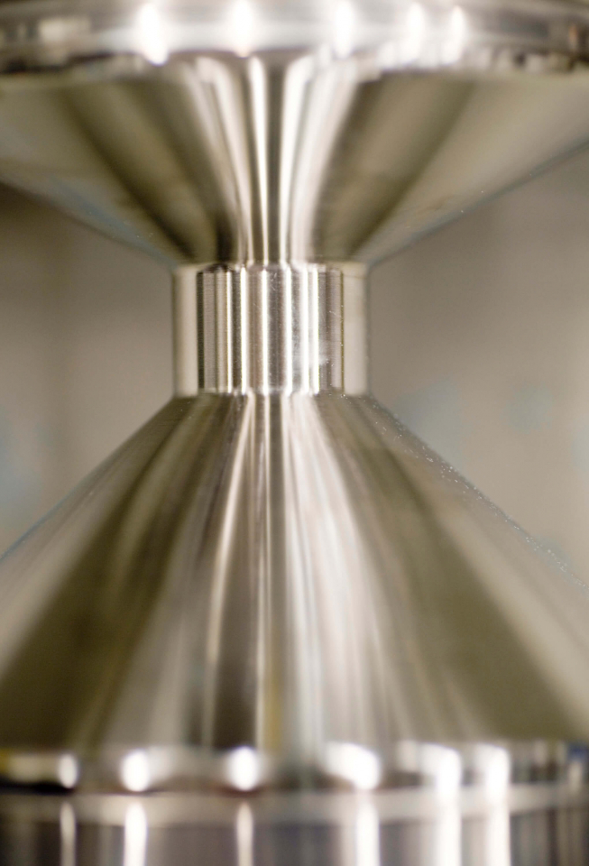
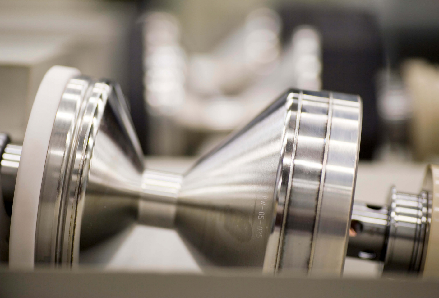
Höchstleistungs-Röntgenstrahler
 Dr. rer. nat. Peter Schardt, Prof. Dr. Dr. rer. nat. Wolfgang Knüpfer, Dr. rer. nat. Karin Söldner")
(f.l.t.r.) Dr. rer. nat. Peter Schardt, Prof. Dr. Dr. rer. nat. Wolfgang Knüpfer, Dr. rer. nat. Karin Söldner
The innovative X-ray tubes developed by Peter Schardt, Karin Söldner, and Wolfgang Knüpfer form the basis. Peter Schardt heads the X-ray and Vacuum Innovation department at Siemens AG in Erlangen, Karin Söldner is project manager and responsible for Straton, Wolfgang Knüpfer is division manager for vacuum technology for X-ray tubes and image intensifiers.
Rapid rotation for sharp pictures
In computer tomography (CT), cross-sectional pictures of the body are taken using x-rays and joined to produce a 3D image, for example, of the heart. To obtain such detailed pictures of a beating heart, the x-ray source, an x-ray tube, must rotate very rapidly around the patient’s body. In this way, it produces many individual pictures with a very short exposure time. The technology used in conventional CT scanners, however, has come up against limiting factors.
more details
Resumes
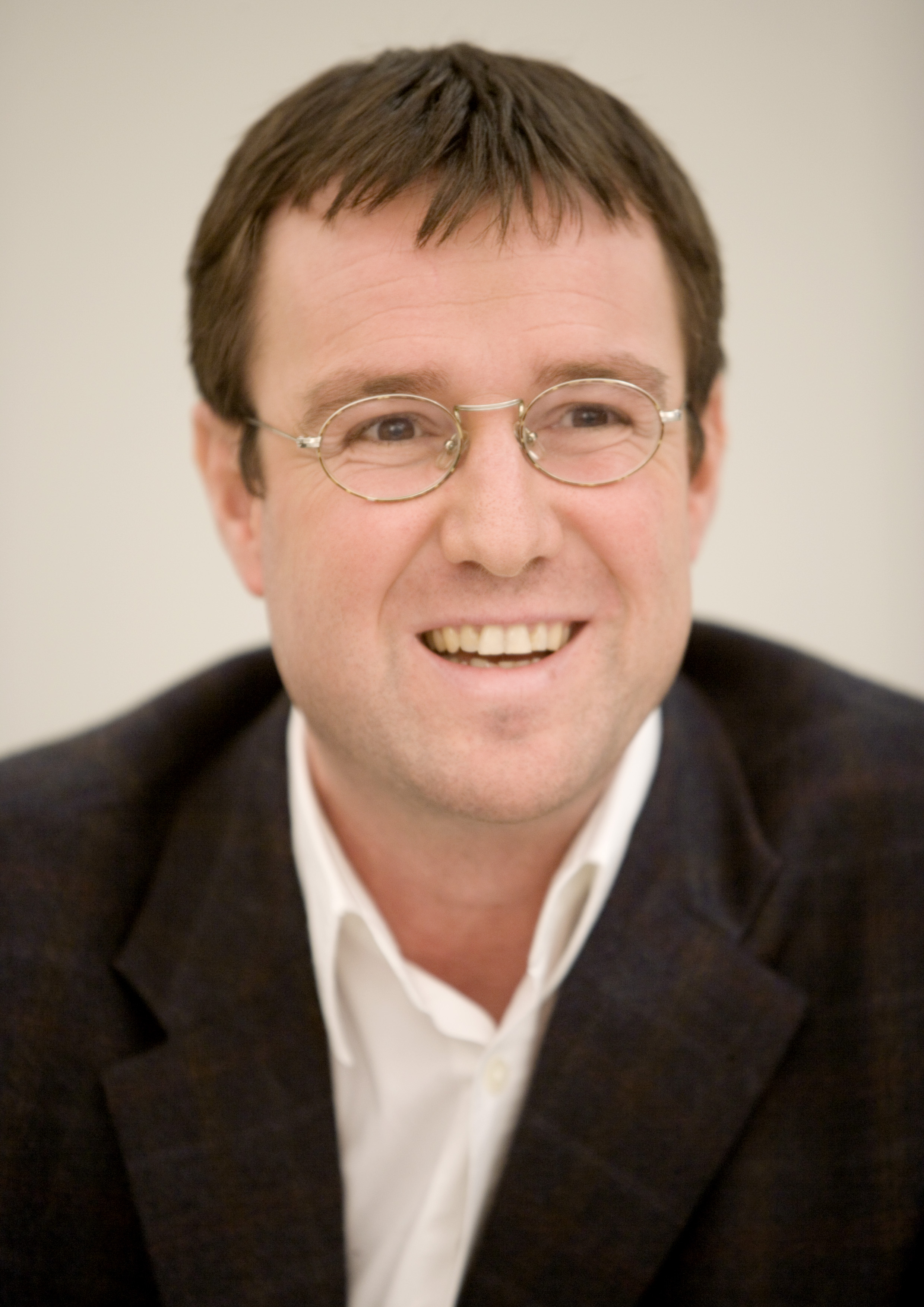
Dr. rer. nat. Peter Schardt
- 20.06.1965
- geboren in Hadamar/Limburg-Weilburg
- 1985 – 1990
- Studium der Physik an der Technischen Universität
Darmstadt - 1990 – 1995
- Doktorand am Institut für Kernphysik,
Technische Universität Darmstadt - 1995
- Promotion
- 1995 – 1996
- Projektleitung für „Neue Röhrenprinzipien“ bei der
Siemens AG, Medical Solutions, Erlangen
- 1996 – 2000
- Projektleitung „Drehkolbenröhre für die medizinische
Diagnostik“ bei der Siemens AG, Medical Solutions,
Erlangen - 2000 – 2002
- Gruppenleiter Vorfeldentwicklung Röntgenröhren bei der
Siemens AG, Medical Solutions, Erlangen - seit 2002
- Abteilungsleitung Vorfeldentwicklung
Röntgen- und Vakuumtechnik bei der Siemens AG,
Medical Solutions, Erlangen
Ehrungen:
- 1995
- Preis für hervorragende wissenschaftliche Leistungen des
Vereins der Freunde der Technischen Universität
Darmstadt e.V. - 2004
- Siemens Medical Innovation Excellence Award 2004
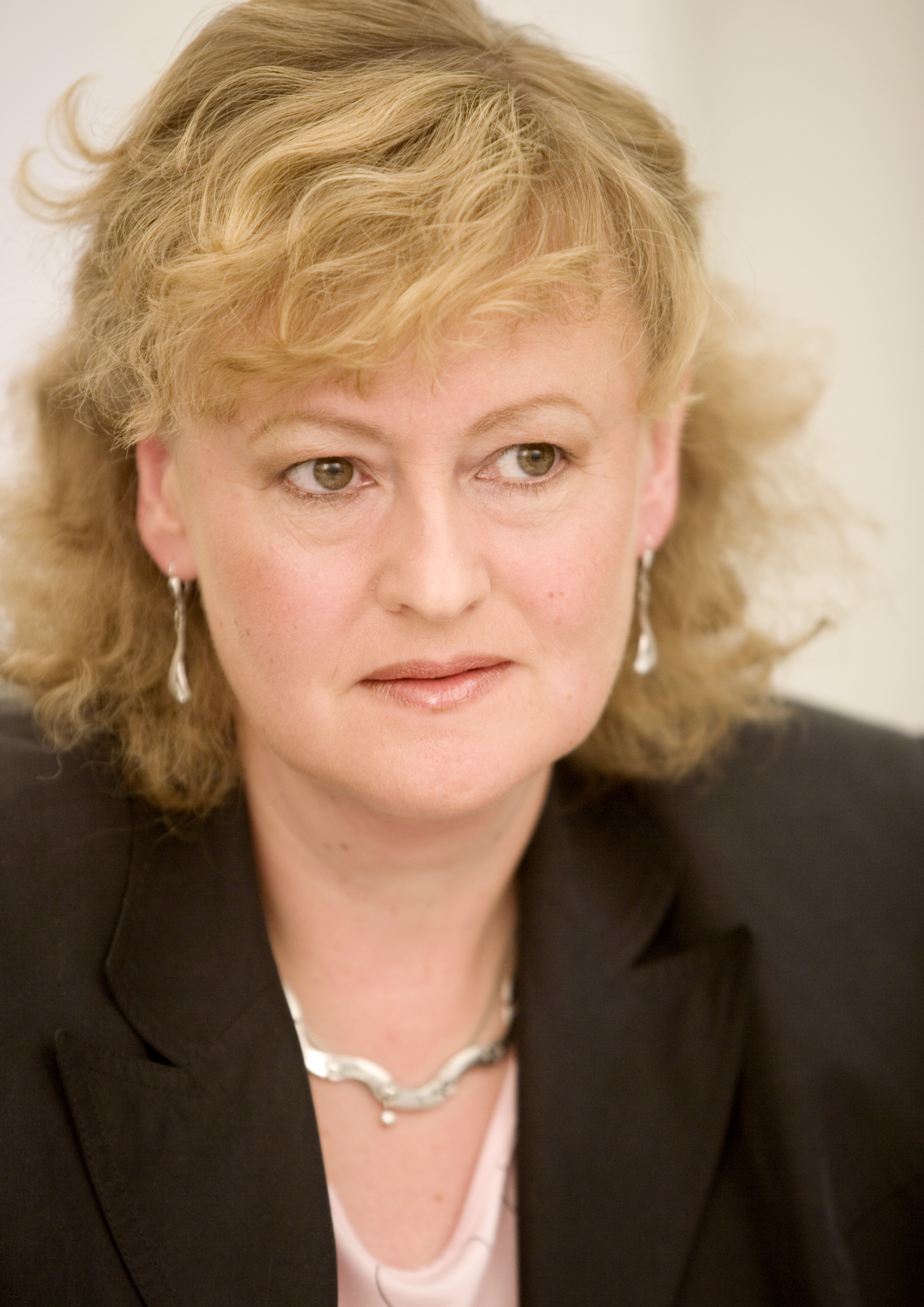
Dr. rer. nat. Karin Söldner
- 06.05.1958
- geboren in Nürnberg
- 1976 – 1982
- Studium der Physik an der Friedrich-Alexander-Universität
Erlangen-Nürnberg - 1982 – 1988
- Doktorand am Physikalischen Institut der Friedrich-
Alexander-Universität Erlangen-Nürnberg - 1987
- Promotion
- 1988 – 1989
- Berechnung von Neutronenflüssen bei der Siemens AG,
Kraftwerk Union (KWU) - 1989 – 1992
- Entwicklung Kugellagertechnologie und Gleitlager-
technologie, Siemens AG, Medical Solutions, RV, Erlangen
- 1992 – 1993
- Mutterschaftsurlaub
- 1993 – 1995
- Mitarbeit im Projekt Gleitlagertechnologie, Betreuung
Fertigungsverlagerung Kugellager zu externer Firma,
Validierungen Kugellagertechnologie
Siemens AG, Medical Solutions, RV, Erlangen - 1995 – 1996
- Projektleitung Projekt Kugellagertechnologie, Qualitäts-
verbesserung und Koordinierung der Aktivitäten in
3 Fertigungslinien Erlangen H, Erlangen TZR und SRW
Siemens AG, Medical Solutions, RV, Erlangen - 1996 – 1999
- Projektleitung bei RV des Förderprojekts der bayerischen
Staatsregierung: Optimierung der Schmierung durch
PVD-Schichten, Kooperation mit GMN, KTmfK der
Universität Erlangen-Nürnberg und MuK der TH Darmstadt,
Entwicklung neuer Lagersysteme für zwei unterschiedliche
CT-Strahler, Siemens AG, Medical Solutions, RV, Erlangen - 1997 – 1998
- Produktverantwortung für CT-Strahler Dura S
Siemens AG, Medical Solutions, RV, Erlangen - 1998 – 1999
- Produktverantwortung für Angiographie-Strahlerfamilie
Megalix
Siemens AG, Medical Solutions, RV, Erlangen - 1999 – 2002
- Mitarbeit im Projekt Diabolo, Koordination von
Konstruktion, Entwicklung, Designverifizierung und
Fertigung bei RVL, Produktverantwortung für Diabolo
Entwicklung Drehkolbenstrahler bis einschließlich
Kundenerprobung
Siemens AG, Medical Solutions, RV, Erlangen - seit 2002
- Projektleiterin und Produktverantwortliche für Straton
Siemens AG, Medical Solutions, RV, Erlangen
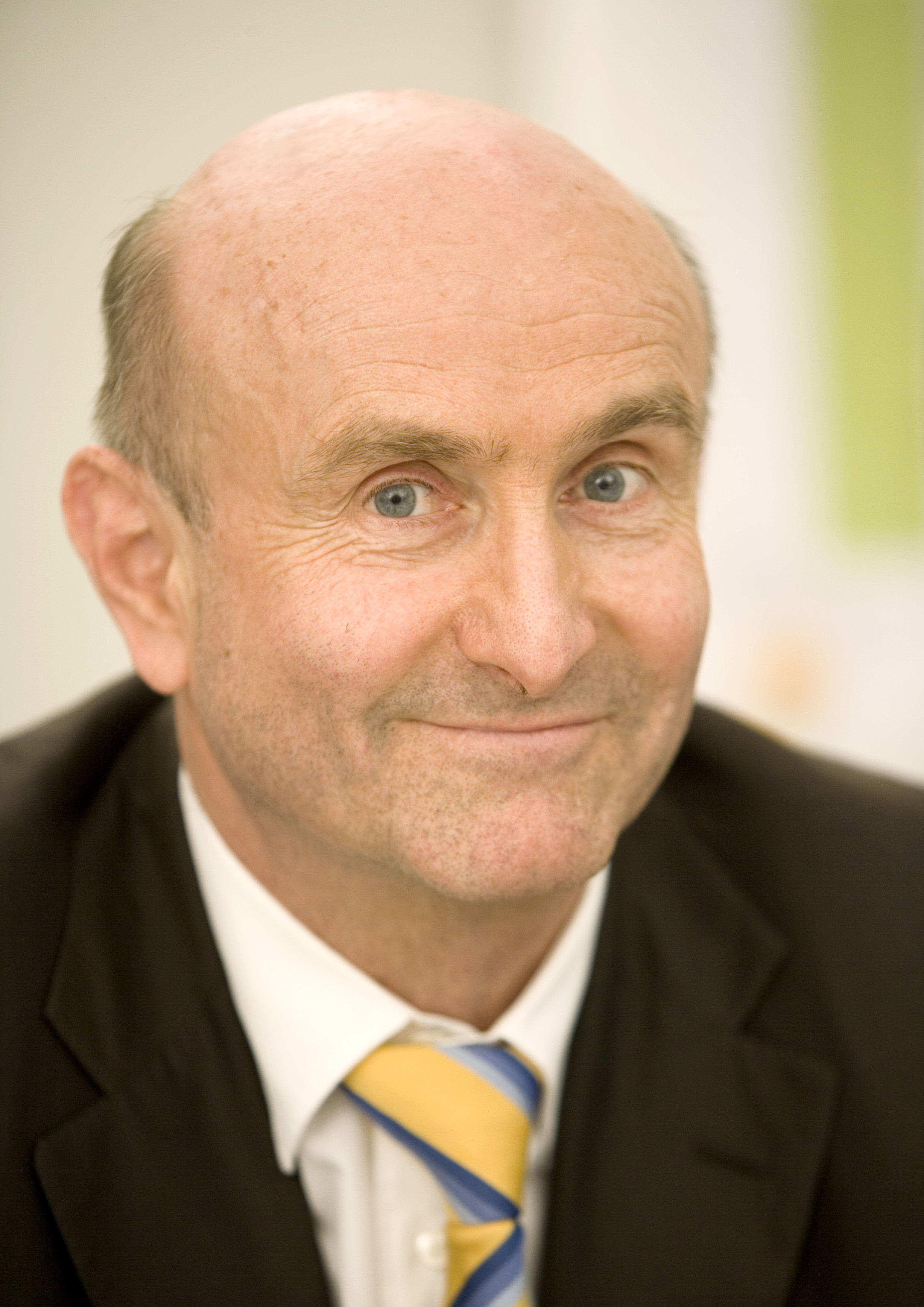
Prof. Dr. Dr. rer. nat. Wolfgang Knüpfer
- 10.04.1943
- geboren in Dresden
- 1962 – 1969
- Studium der Physik an der Friedrich-Alexander-Universität
Erlangen-Nürnberg - 1969 – 1972
- Doktorand am Institut für Theoretische Physik,
Friedrich-Alexander-Universität Erlangen-Nürnberg - 1972
- Promotion
- 1972 – 1975
- Assistent am Institut für Theoretische Physik,
Friedrich-Alexander-Universität Erlangen-Nürnberg
Aufbau einer Forschungsgruppe mit Projekten in
Kern- (Kernstruktur) und Strahlenphysik
International anerkannte Zusammenarbeit mit dem Institut
für Kernphysik der TH Darmstadt (S-DALINAC)
- 1975 – 1981
- Forschungsaufenthalte in Washington, Paris, Utrecht
- 1981 – 1983
- Habilitation, Privatdozent
- 1983 – 1985
- Assistent der Entwicklungsleitung Röntgen der
Siemens AG, Medical Solutions, Erlangen - 1985 – 1989
- Entwicklungsleiter X-Ray Speicherleuchtstoffe
und Verstärkerfolien
Siemens AG, Medical Solutions, Erlangen - 1989 – 1992
- Entwicklungsleiter Röntgenbildverstärker
Siemens AG, Medical Solutions, Erlangen - 1992
- Geschäftszweigleiter Bildverstärker
Siemens AG, Medical Solutions, Erlangen - seit 1993
- Geschäftsgebietsleiter Vakuumtechnik
(Entwicklung, Produktion, Marketing/Vertrieb)
für Röntgenröhren und Bildverstärker
Siemens AG, Medical Solutions, Erlangen - 1994
- apl. Professor für Physik an der Friedrich-Alexander-
Universität Erlangen-Nürnberg - seit 1992
- Mitglied der AAPM (American Association of Physicists in
Medicine) - 1997 – 2002
- Mitglied des Wissenschaftlichen Rates der GSI (Gesellschaft
für Schwerionenforschung), Darmstadt
Contact
Spokesperson
Dr. rer. nat. Peter Schardt
Abteilungsleiter Grundlagen / Vorfeldentwicklung
Vacuum Technology (RV)
Siemens AG, Medical Solutions
Günther-Scharowsky-Str. 1
91058 Erlangen
Tel.: +49 (0) 9131 / 73 23 70
Fax: +49 (0) 9131 / 73 24 27
E-Mail: peter.schardt@siemens.com
Press
Axel Wieczorek
Leiter Public Communications
Corporate Communications
Siemens AG, Medical Solutions
Henkestr. 127
91052 Erlangen
Tel.: +49 (0) 9131 / 84 83 35
Fax: +49 (0) 9131 / 84 29 24
E-Mail: a.wieczorek@siemens.com
A description provided by the institutes and companies regarding their nominated projects
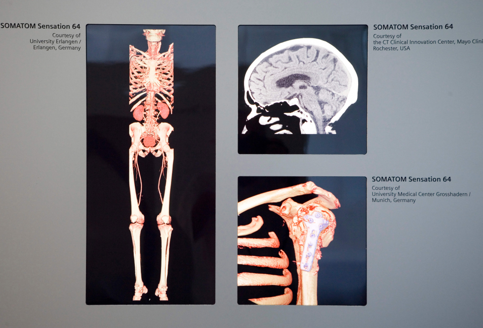
Cardiovascular diseases are the number one cause of death in Germany. In order to detect diseased coronary vessels early, CT scanners are being used every more frequently.
But how do these machines produce the necessary degree of precision?
The innovative X-ray tubes developed by Peter Schardt, Karin Söldner, and Wolfgang Knüpfer form the basis. Peter Schardt heads the X-ray and Vacuum Innovation department at Siemens AG in Erlangen, Karin Söldner is project manager and responsible for Straton, Wolfgang Knüpfer is division manager for vacuum technology for X-ray tubes and image intensifiers.
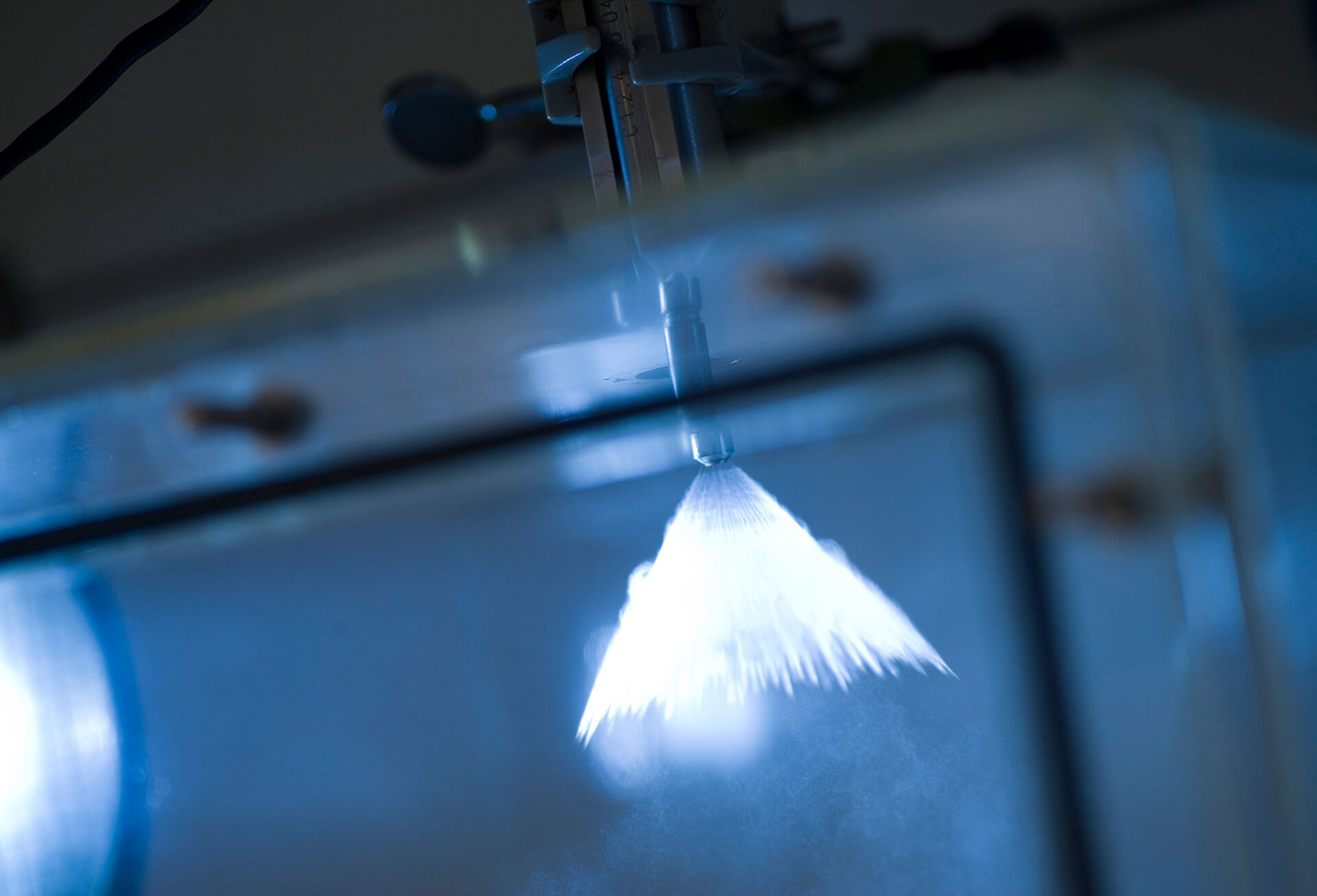
Rapid rotation for sharp pictures
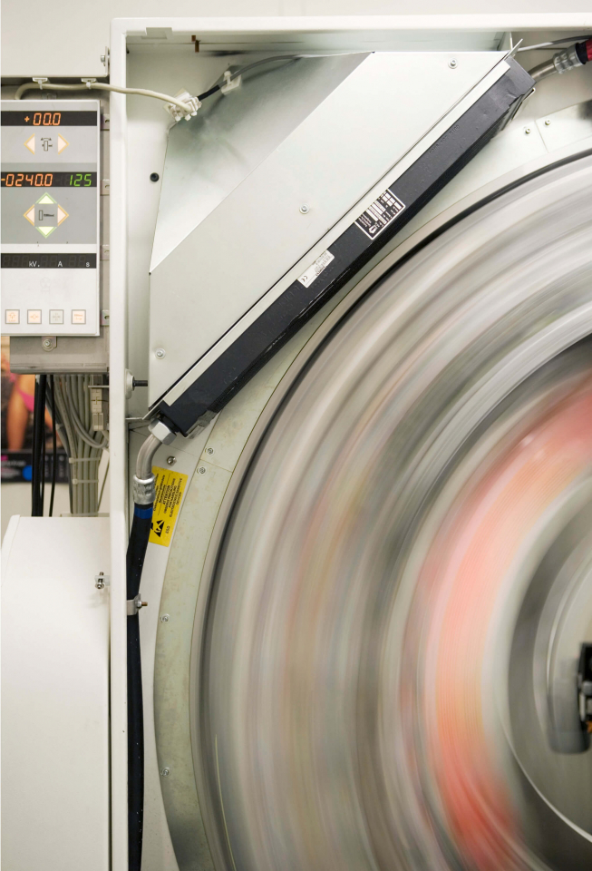
In computer tomography (CT), cross-sectional pictures of the body are taken using x-rays and joined to produce a 3D image, for example, of the heart. To obtain such detailed pictures of a beating heart, the x-ray source, an x-ray tube, must rotate very rapidly around the patient’s body. In this way, it produces many individual pictures with a very short exposure time. The technology used in conventional CT scanners, however, has come up against limiting factors.
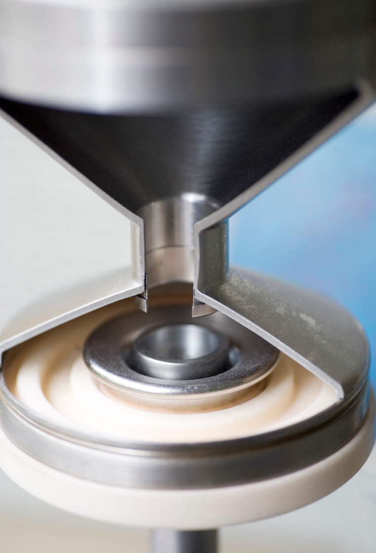
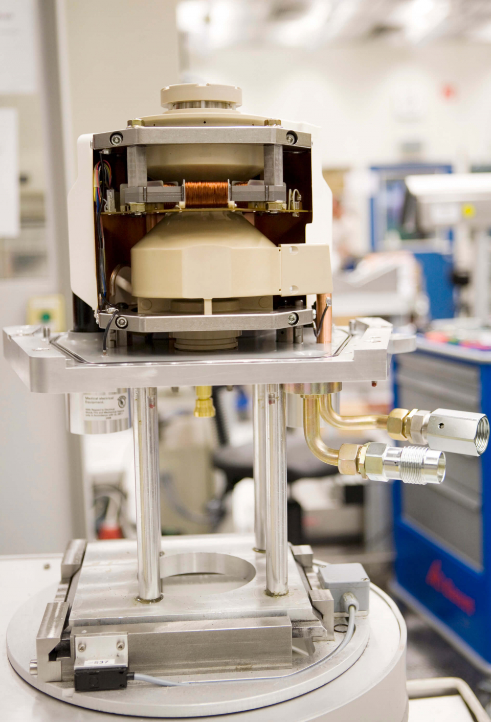
To produce x-rays in the tube anodes, vast amounts of heat are produced. To cool these down fast enough, the x-ray tubes have to be large and heavy. This limits the rate of rotation. The “Straton” x-ray tube technology developed by the three nominees makes it possible to build smaller and yet more powerful x-ray tubes – thanks to the innovative technology in the tubes.
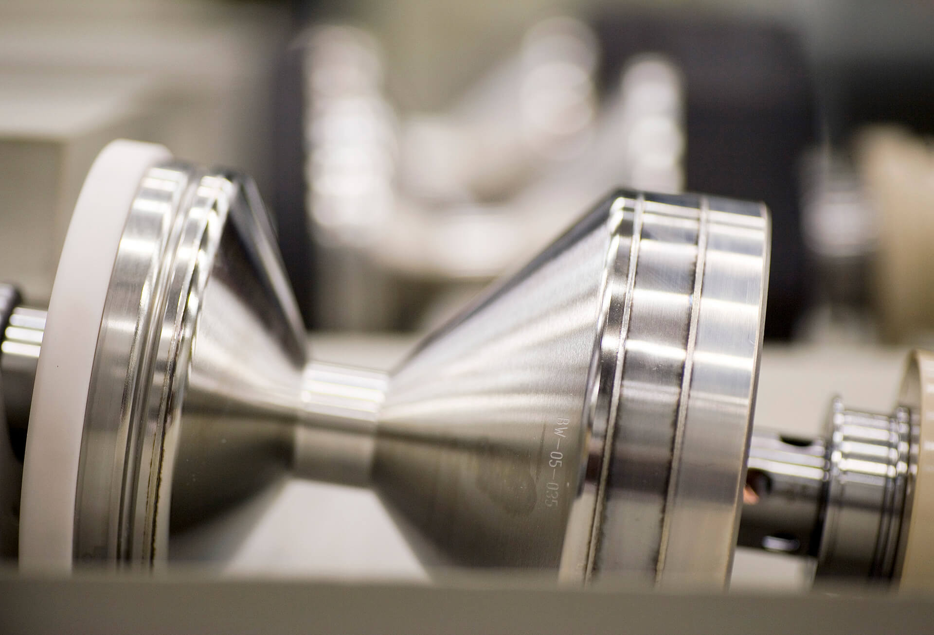
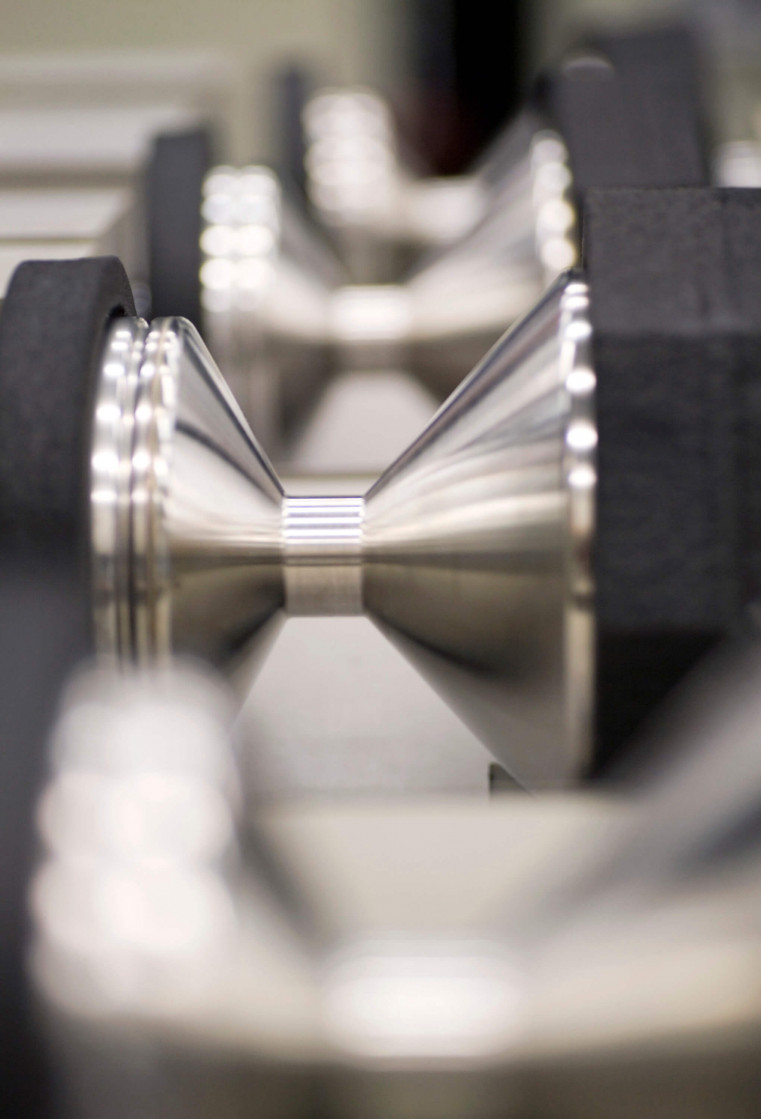
The entire tube turns
Instead of an anode that turns inside the tube but independent of it, the entire x-ray tube rotates rapidly about its axis. The anode is rigidly attached to it and is directly cooled by an oil circuit. The advantage: the x-ray tube design can be built very compact – and cools down very rapidly. The result is a very accurate look inside the human body – with considerably shorter examination times. The coronary arteries and blood vessels can be imaged in brilliant quality with a single CT scan lasting no more than a few seconds. And: patents can be examined despite a rapid or irregular heartbeat – something conventional tomographs can’t do.
With the new, fast x-ray tubes, however, the heartbeat can be virtually frozen on the images. The Straton technology is now used exclusively in the latest generation of Siemens computer tomographs which were introduced in 2003. These scanners have also improved emergency diagnostics: the physician can produce a whole body scan in around 20 seconds in order to obtain a picture of an accident victim’s internal injuries.
The right to nominate outstanding achievements for the German Future Award is incumbent on leading German institutions in Science and Industry as well as foundations.
The Project „Revolutionary high-speed, high-resolution X-ray tube for computer tomography“ was nominated by the Verband Technisch-Wissenschaftlicher Vereine.