 Gebärdensprache
 Leichte Sprache

Nominee 2019
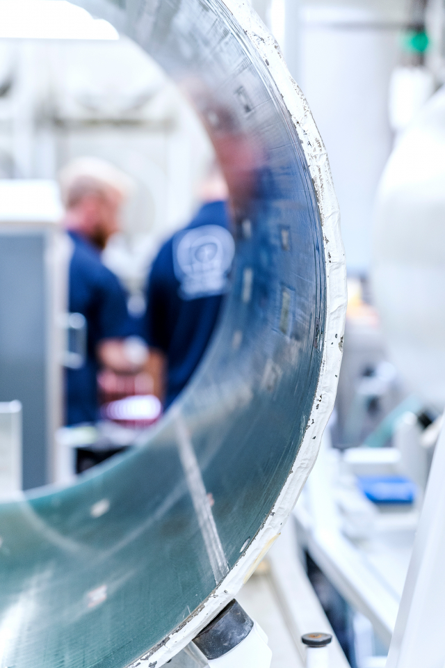
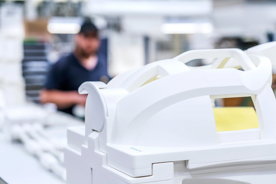
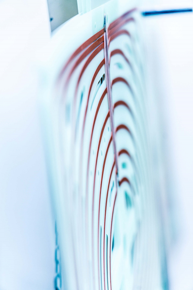
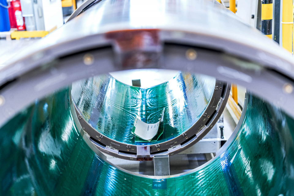
Ultra-high-field MRI
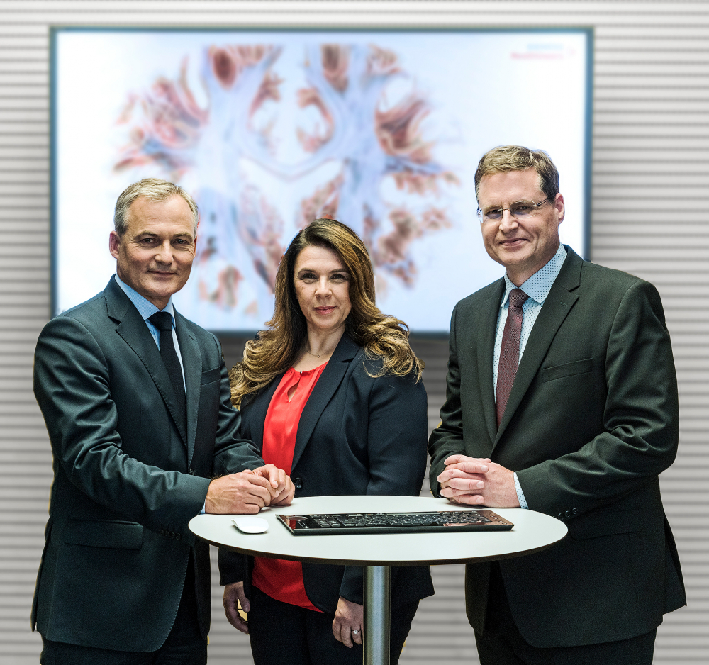
(f.l.t.r.) Univ.-Prof. Dr. med. Arnd Dörfler, Christina Triantafyllou, Ph.D., Univ.-Prof. Dr. sc. techn. Mark E. Ladd
Christina Triantafyllou, PH.D., Univ.-Prof. Dr. med. Arnd Dörfler and Univ.-Prof. Dr. sc. techn. Mark E. Ladd have the answer. The three nominees have considerably increased the strength of the magnetic field used in MRI - and are providing new and more detailed insights into the human body. By taking innovative approaches, they have succeeded in making the advanced ultra-high-field MRI technology accessible not only for basic research, but now also for use in hospitals.
Christina Triantafyllou heads the Global Ultra-High-Field Solutions team at Siemens Healthineers AG in Erlangen, Arnd Dörfler is head of the Neuroradiology department at Universitätsklinikum Erlangen of Friedrich-Alexander University Erlangen-Nürnberg and Mark E. Ladd is head of the department of Medical Physics in Radiology at the German Cancer Research Center, Deutschen Krebsforschungszentrum, Heidelberg.
More Details
Resume
Christina Triantafyllou, Ph.D.
- 15.7.1970
- Born in Stuttgart, Germany
- 1988
- Apolytirio, High School Diploma Eniaio Polycladiko Senior High School, Katerini, Pierias, Greece
- 1994
- B.Sc., Physics Aristotle University of Thessaloniki, Thessaloniki, Greece
- 1995
- M.Sc., Medical Physics University of Surrey, Guildford, UK
- 1997 – 2001
- Ph.D., Medical Physics Institute of Psychiatry, Guy’s, King’s and St Thomas’ School of Medicine, King’s College, University of London, London, UK
- 2001 - 2006
- Harvard University, A.A. Martinos Center for Biomedical Imaging,
MRI Physics Group, Department of Radiology, Massachusetts General Hospital, Harvard Medical School Boston, MA, USA
Postdoctoral Research Fellow - 2006 – 2014
- Harvard University, Department of Radiology, Massachusetts General Hospital, Harvard Medical School
Boston, MA, USA Assistant in Neuroscience - 2006 – 2014
- Harvard University, A.A. Martinos Center for Biomedical Imaging, Department of Radiology, Massachusetts General Hospital, Harvard Medical School
Boston, MA, USA Faculty, Instructor - 2006 – 2012
- Massachusetts Institute of Technology (MIT), Department of Brain and Cognitive Sciences, A.A. Martinos Imaging Center, McGovern Institute for Brain Research
Cambridge, MA, USA Head MRI Physicist - 2006 – 2012
- Massachusetts Institute of Technology (MIT), Department of Brain and Cognitive Sciences, A.A. Martinos Imaging Center, McGovern Institute for Brain Research
Cambridge, MA, USA Research Scientist - 2012 – 2012
- Massachusetts Institute of Technology (MIT), Department of Brain and Cognitive Sciences, A.A. Martinos Imaging Center, McGovern Institute for Brain Research
Cambridge, MA, USA Principal Research Scientist - 2007 – 2012
- Massachusetts Institute of Technology (MIT), Department of Brain and Cognitive Sciences, A.A. Martinos Imaging Center, McGovern Institute for Brain Research
Cambridge, MA, USA Associate Director - 2012 – 2013
- Siemens AG, Healthcare Sector, Business Line MRI
Erlangen, Germany
Director of Global Ultra High Field Business Management - 2013 – 2018
- Siemens Healthineers, Business Line MR
Erlangen, Germany
Director of Global Ultra High Field Product Relationship Management - Since 2018
- Siemens Healthineers, Business Line MR
Erlangen, Germany
Solution Owner, Product Line Research and Clinical Translation - Since 2018
- Siemens Healthineers, Business Line MR Erlangen, Germany
Director of Global Ultra-High Field MR Solutions
Team leader of Solutions and Digital, Product Line Research and Clinical Translation
Further Activities
- 38 Peer reviewed publications in scientific journals
94 scientific abstracts in Conference Proceedings
Co-Author of 2 book chapters
More than 25 invited talks by international scientific societies and universities
Ad Hoc Reviewer for several scientific journals and professional societies
Grant Review Activities (Department of Veterans Affairs, Rehabilitation R&D Study Section, Grant Review Panel, Washington DC, USA, 2011)
Member of professional scientific societies (International Society for Magnetic Resonance in Medicine, European Society of Magnetic Resonance in Medicine, Organization for Human Brain Mapping, Institute of Physics in UK) - Since 2013
- Organizing Committee of the High Field Systems & Applications Study Group, International Society for Magnetic Resonance in Medicine (ISMRM)
- Since 2019
- Member of the group, Women of ISMRM (International Society for Magnetic Resonance in Medicine)
- 2005
- Software Developer, Collaboration Exchange Program, Department of MREA, Advanced Neuro Applications Group, Siemens Healthcare, Erlangen, Germany
- 2000 – 2001
- Research Associate in Medical Image Processing, Robert Steiner MRI Unit, Imaging Sciences Department, Hammersmith Hospital, Imperial College, London, UK
- 1996
- Clinical Scientist, Nuclear Medicine Department, Leicester Royal Infirmary Hospital, Leicester, UK
- 1996
- Student Internship Placement, Nuclear Medicine Department, Hammersmith Hospital, London, UK
- 1993
- Summer School in Research Advanced Physics, Department of Physics, University of Crete, Greece
Honors (selected)
- 2010
- Infinite Mile Award for Outstanding Achievement, Office of the Provost, Office of the Vice President for Research and Associate Provost, Massachusetts Institute of Technology (MIT), Cambridge, MA, USA
- 2010
- Poster Award (1st place), 2nd Annual Scientific Symposium, Ultra High Field Magnetic Resonance: Clinical Needs, Research Promises and Technical Solutions, B.U.F.F., Berlin, Germany
- 1997
- Scholarship Award for Ph.D. studies, The Stroke Association, King’s College, University of London, London, UK
Travel Stipends, International Society for Magnetic Resonance in Medicine, 13th Annual Meeting, Miami, Florida, USA, 2005, 12th Annual Meeting, Kyoto, Japan, 2004, 11th Annual Meeting, Toronto, Canada, 2003, 9th Annual Meeting, Glasgow, Scotland, UK, 2001
Travel Award, Organization for Human Brain Mapping, 6th Annual Meeting, San Antonio, Texas, USA, 2000
Travel Stipends, British Chapter of the International Society for Magnetic Resonance in Medicine, 5th Annual Meeting, London, 1999, 3rd Annual Meeting, Manchester, UK, 1997
Travel Award, European Society for Magnetic Resonance in Medicine & Biology, 15th Annual Meeting, Geneva, Switzerland, 1998
Research Scientific Interests
- Methodological advances in magnetic resonance imaging, including imaging with ultra-high magnetic fields
Ultra high resolution in functional magnetic resonance imaging
Clinical neuro applications with ultra-high field MRI
Univ.-Prof. Dr. med. Arnd Dörfler
- 17.12.1967
- Born in Karlsruhe, Germany
- 1987 - 1994
- Human Medicine Studies at the Universities of Heidelberg and Zurich
- 1994
- Doctorate (Dr. med.)
University of Heidelberg, Department of Neuroradiology
‘Magnetic resonance tomography in the acute phase of cerebral ischemia - animal experiments using two ischemia models’ - 2002
- Habilitation including venia legendi
University of Duisburg-Essen, Essen, Germany
‘Radiological diagnosis and therapy of middle cerebral artery infarction’ - 4.2000
- Board Certification in ‘Diagnostic Radiology’
- 7.2003
- Board Certification in ‘Neuroradiology’
- 2006
- Master’s Degree ‘Management of Health and Social Facilities’
University of Kaiserslautern and Witten/Herdecke, Germany - 12.1994 - 5.1995
- Department of Neuroradiology, University Hospital Heidelberg
- 6.1995 - 5.1996
- Department of Neurology, University Hospital Heidelberg (‘clinical year’)
- 6.1996 - 6.1997
- Department of Neuroradiology, University Hospital Heidelberg
- 7.1997- 11.2004
- Institute for Diagnostic and Interventional Radiology and Neuroradiology, Essen University Hospital
- since 12.2004
- Head of Department of Neuroradiology, University Hospital Erlangen
Professorship (C3, Extraordinarius) of Neuroradiology at Friedrich-Alexander-University, Erlangen-Nuremberg - since 8.2014
- Professor (W3, Ordinarius) of Neuroradiology at Friedrich-Alexander-University, Erlangen-Nuremberg
- 2013
- Offer of an appointment as full professor (W3) of Neuroradiology at the Charité, University Medicine Berlin, declined after being offered a W3 professorship at Friedrich-Alexander-University, Erlangen-Nuremberg in 7/2014
- 5/2016
- Offer of an appointment as full professor of Neuroradiology at the University of Zurich, in conjunction with the directorship of the Medical Center of Neuroradiology at University Hospital Zurich (declined in 10/2016)
Further activities
- More than 300 publications in scientific journals
Author of 17 book chapters and 24 review articles
Expert reviewer for more than 10 scientific journals and a number of national (e.g. DFG) and international funding organizations (e.g. SNF, NIH) and societies - Since 2006
- Extended board member of the German Society for Neuroradiology (DGNR)
- Since 2014
- Full board member of the German Society for Neuroradiology (DGNR)
- 2016 - 2018
- President of the German Society for Neuroradiology (DGNR)
- Since 2019
- Board Member of the German Radiological Society (DRG)
Past-President of the German Society for Neuroradiology (DGNR)
Research Interests
Honors (selected)
- 1998
- “Certificate of Excellence“ Symposium Neuroradiologicum, Philadelphia, USA
- 2002
- Kurt-Decker Award of the German Society for Neuroradiology
- 2006
- Felix-Wachsmann Award of the Academy for education and training of the German Radiological Society
- 2010
- ‘Best in science’ Award, Society for Vascular and Interventional Neurology, San Francisco, USA
- 2013
- Glaucoma Research Award, German Ophthalmological Society
Univ.-Prof. Dr. sc. techn. Mark E. Ladd
- 7.9.1967
- Born in Wayne, Michigan, USA
- 1982 – 1985
- High School Diploma John Glenn High School, Westland, Michigan, USA
- 1985 – 1989
- Bachelor of Science in Engineering University of Michigan, Ann Arbor, USA
- 1989 – 1991
- Master of Science in Electrical Engineering Stanford University, Stanford, California, USA
- 1995 – 1998
- PhD (Dr. sc. techn.) Swiss Federal Institute of Technology (ETH) Zurich, Switzerland
- 2001
- Habilitation (post-doctoral thesis) with venia legendi University of Duisburg-Essen, Essen, Germany
- 1986, 1987
- General Motors Corporation, Warren, Michigan USA Advanced Manufacturing Engineering Staff Engineering Intern
- 1989 – 1991
- Stanford University, Stanford, California, USA Space, Telecommunications and Radioscience Laboratory Research Assistant
- 1992 – 1994
- General Electric Medical Systems, Milwaukee, Wisconsin USA Engineer
- 1994 – 1999
- General Electric Medical Systems in cooperation with University Hospital Zurich, Switzerland Advanced Systems Engineer
- 1999 – 2004
- University Hospital Essen, Germany Department of Diagnostic and Interventional Radiology and Neuroradiology Senior Physicist
- 2000 – 2010
- MR-Innovation GmbH, Essen, Germany Vice President Research and Development and Safety Officer
- 2004 – 2013
- University Hospital Essen, Germany Department of Diagnostic and Interventional Radiology and Neuroradiology Professor of Biomedical Imaging
- 2006 – 2013
- University of Duisburg-Essen, Essen, Germany Erwin L. Hahn Institute for Magnetic Resonance Imaging Director
- Since 2013
- University of Duisburg-Essen, Essen, Germany Erwin L. Hahn Institute for Magnetic Resonance Imaging Principle Investigator
- Since 2013
- University of Heidelberg, Germany Medical Faculty Professor for Medical Physics in Radiodiagnostics and Biophysics
- Since 2013
- German Cancer Research Center (DKFZ), Heidelberg, Germany Division of Medical Physics in Radiology Head
Further Activities
- More than 260 publications in scientific journals
Author of 11 book chapters and 20 review articles
Reviewer for more than 20 scientific journals and a variety of national and international funding organizations and professional societies - 2010 – 2013
- Board of Trustees, International Society for Magnetic Resonance in Medicine (ISMRM)
- 2012 – 2018
- Deputy Editor of „Magnetic Resonance in Medicine”
- 2013
- Guest Editor of „Investigative Radiology”, Special Issue on Clinical Advances with 7 Tesla
- 2013 – 2017
- Governing Committee of the High Field Systems & Applications Study Group, International Society for Magnetic Resonance in Medicine (ISMRM)
- Since 2013
- Scientific Advisory Board of „Der Radiologe“
- 2016
- Co-Chair of Workshop „UHF MRI: Technological Advances & Clinical Applications”, International Society for Magnetic Resonance in Medicine (ISMRM)
- 2017 – 2018
- Vice President of the German Society for Medical Physics (DGMP)
- Since 2019
- President of the German Society for Medical Physics (DGMP)
Research Interests
- Methodological advances in magnetic resonance imaging and spectroscopy, including imaging with ultra-high magnetic fields, parallel transmission, MRI safety, and magnetic resonance-guided radiotherapy
Honors (selected)
- 2000
- Award winner in the Competition for the Promotion of Young Academicians as part of the Program for Research Innovation of the State of North Rhine-Westphalia, Germany
- 2006
- Award winner in the Innovation Competition for the Promotion of Medical Engineering of the German Federal Ministry of Education and Research (BMBF)
- 2012
- Recipient of an Advanced Grant from the European Research Council (ERC)
- Multimodal imaging approaches of neurological disease
7 Tesla high-field MR neuroimaging
Interventional neuroradiology
Contact
Press
Ulrich Künzel
Communications, Media Relations
Siemens Healthcare GmbH
Karlheinz-Kaske-Str. 5
91052 Erlangen
Phone: +49 (0) 162 / 24 33 492
E-Mail: ulrich.kuenzel@siemens-healthineers.com
Spokesperson
Christina Triantafyllou,Ph.D.
Siemens Healthcare GmbH
Allee am Röthelheimpark 2
91052 Erlangen
Phone: +49 (0) 9131 / 84 54 94
E-Mail: christina.triantafyllou@siemens-healthineers.com
A description provided by the institutes and companies regarding their nominated projects
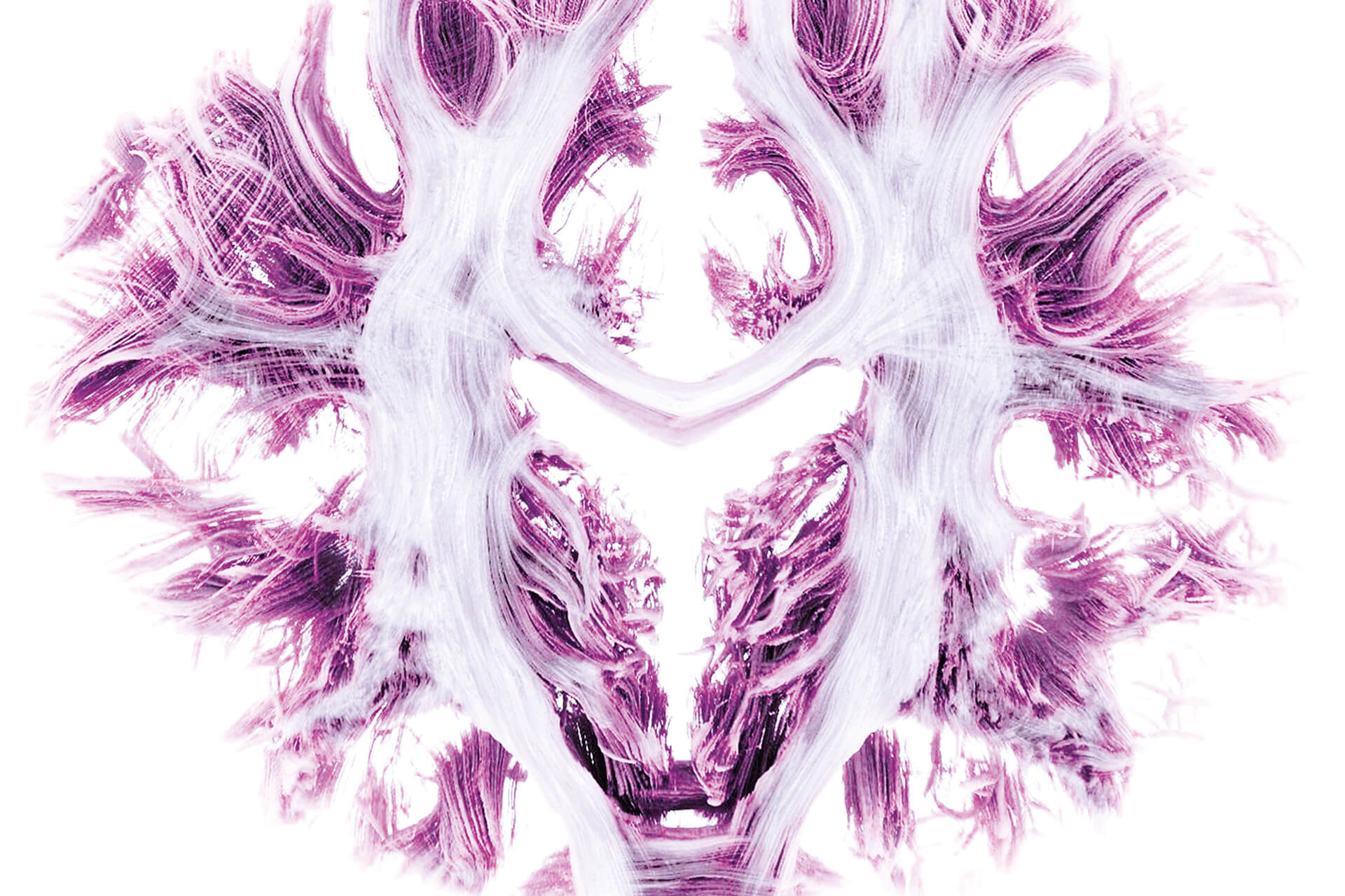
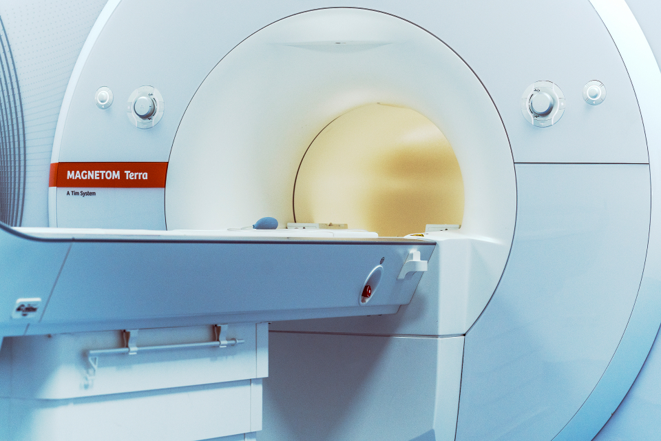
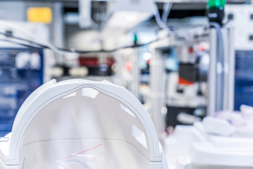
Magnetic resonance imaging (MRI) – also known as magnetic resonance tomography – is now the leading diagnostic imaging method and plays a crucial role in the diagnosis of a large number of diseases. With the development of Siemens Healthineers’ Magnetom Terra, the first ultra-high-field MRI system for clinical use, Christina Triantafyllou Ph.D., Prof. Mark E. Ladd and Univ.-Prof. Arnd Dörfler achieved a breakthrough in diagnostic imaging and at the same time established 7 Tesla as a new clinical field strength. The new system is the first in the world to enable the use of ultra-high-field MRI in clinical applications. The research and development team has thus broken new grounds in medical imaging and reached a significant milestone in the history of MRI.
In particular, for diseases of the central nervous system, internal organs, blood vessels and musculoskeletal system, 7-Tesla MRI offers precise visualisation of the smallest structures in the human body and thus often the best possible diagnosis. Particularly in the early stages of diseases such as multiple sclerosis (MS) and neurodegenerative diseases, for example dementia and Parkinson's disease, pathological changes are often not sufficiently pronounced to be diagnosed using the clinically established MRI systems at lower field strengths. The image quality and spatial resolution achievable using conventional MRI systems are not always adequate for this purpose. Consequently, before the diagnosis is achieved or the treatment is started, valuable time might be lost. The Magnetom Terra from Siemens Healthineers promises a fundamental improvement here: Magnetom Terra’s ultra-high-field imaging provides a very high level of detail and is thus able to reveal the slightest changes in the anatomy and even in the function of organs. This helps to enable firstly very early diagnosis and secondly reliable differentiation from other diseases, something which was not achievable with previous clinically established field strengths.
In 2017, with Magnetom Terra, the German-Future-Prize-nominated research and development team made possible that ultra-high-field MRI with a field strength of 7 Tesla, which has only been utilized in basic research since the early 2000s, became available for clinical use worldwide, for the first time, as a new and effective diagnostic method – consequently also benefiting patients directly. Christina Triantafyllou leads the global team ‘Ultra-High-Field MRI Solutions’ at Siemens Healthineers, Mark E. Ladd is head of the Division of Medical Physics in Radiology of the German Cancer Research Center in Heidelberg and Arnd Dörfler heads the Neuroradiological Department at the University Hospital of the Friedrich-Alexander University of Erlangen-Nuremberg.
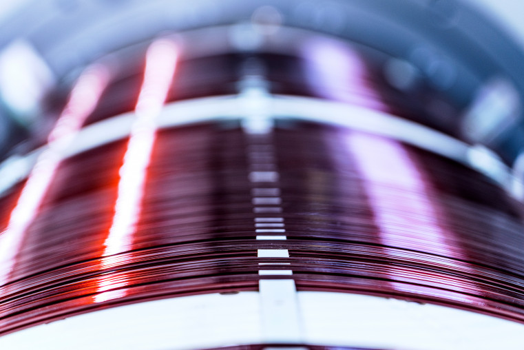
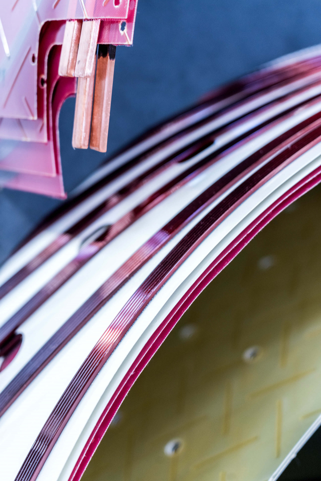
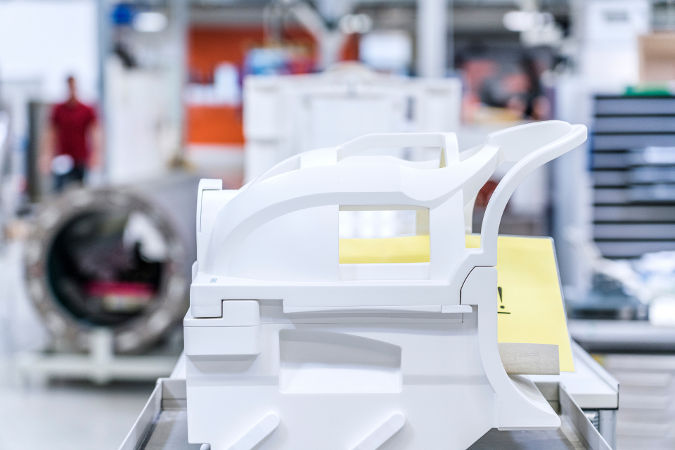
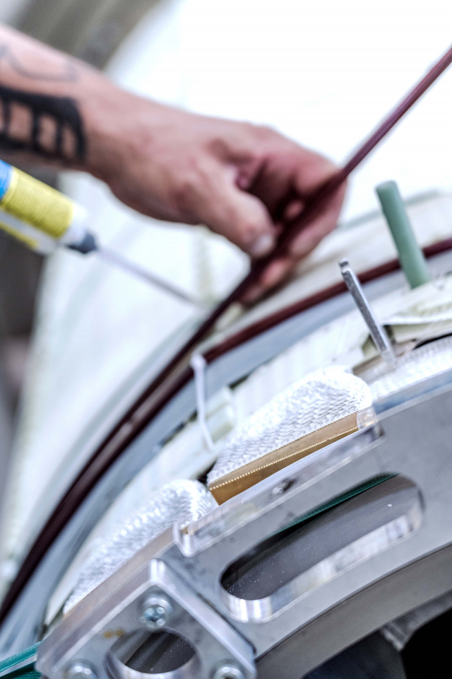
Besides making ultra-high-field MRI at 7 Tesla available for use in a clinical environment, Magnetom Terra also represents an engineering accomplishment that had not previously been achieved: not only new safety concepts have been designed for use on humans, but also a brand new designed, innovative actively-shielded magnet has been developed, which is only half as heavy as the magnets used in previous research systems. Magnetom Terra is therefore much easier to transport and, in particular, easier to integrate into existing hospital infrastructure. Time-consuming and costly construction of new buildings can thus be avoided. A further innovation is Magnetom Terra’s dual-mode functionality, which enables users to switch directly between clinical and research use. The system thus offers the ideal platform for translational research – the latest research developments can therefore be used directly for the benefit of patients.
Early diagnosis and more effective countermeasures to combat multiple sclerosis
The innovative metabolic imaging technology of Magnetom Terra can render pathological changes visible at the metabolic level, even before they manifest themselves either morphologically and anatomically. Here, the field strength of 7 Tesla, which is equivalent to approximately 140,000 times the strength of the earth’s magnetic field, produces impressive results. While it was previously only possible to detect pathological changes on clinical images only at the advanced stages of MS, for example at 7 Tesla these can often be identified in earlier stage of the disease. Furthermore, smaller lesions in the brain’s gray matter can be detected, which previously was not possible. Subsequently, such early diagnosis and prompt initial treatment are of particular importance for MS patients, many of whom are young, in delaying the onset of disability or even preventing it entirely as the disease progresses.[1], [2]
Precise identification of epileptogenic foci
Epilepsy is one of the most common neurological conditions. In Germany alone, there are around half a million epilepsy patients. Even given the best drug treatment, a considerable proportion of these patients do not remain seizure-free, which greatly restricts the lives of those affected. On the other hand, this patient group may benefit from a higher success rate from epilepsy surgery. This intervention involves the surgical removal of epileptogenic foci from the patient’s brain. After complete removal, patients can live seizure-free. Magnetom Terra’s outstanding detail resolution makes it possible for epileptogenic foci to be diagnosed more precisely or even initially identified as such. This makes interventions more reliable, enabling those affected to regain their original quality of life.[3], [4]
Sodium imaging for neurodegenerative diseases
With dementia disorders, a diagnosis can generally be made only in the advanced stages. Morphological damage in the brain will then already have occurred. Here, too, Magnetom Terra offers new possibilities for detecting Parkinson’s disease or Alzheimer’s disease at an early stage. Using ultra-high-field MRI, the deposits typical of Alzheimer’s can be shown better than previously, and in the case of Parkinson’s, structural changes in very specific areas of the brain can reliably be detected at an early stage. Here, anatomically visible features play as much a role [5] as metabolic changes, which can be shown clearly for example by sodium imaging.[6] The release of Magnetom Terra’s sodium-based metabolic MRI imaging system for clinical use in Europe and the USA in 2018 was a worlds first.
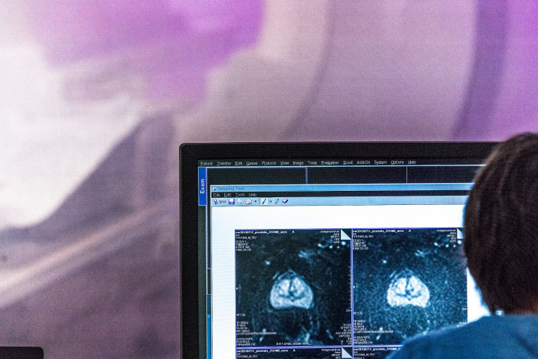
Improved treatment monitoring in oncology
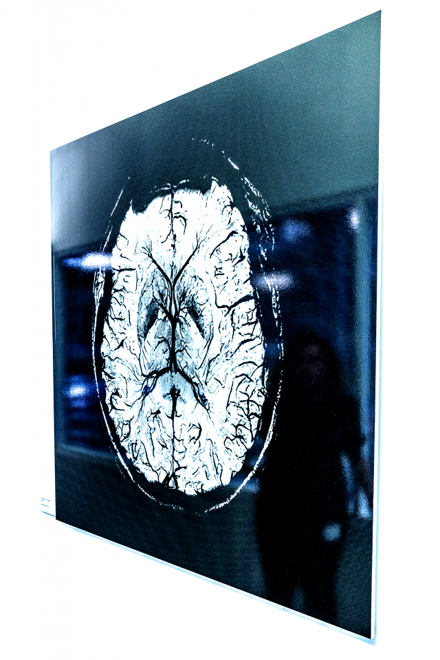
In oncology, Magnetom Terra also offers new opportunities for early treatment and better monitoring. While cancers can frequently be diagnosed using previously clinically established field strengths, weeks can often pass until it is clear whether a treatment has been effective. Using biomarkers, ultra-high-field MRI has succeeded in clinical research in characterizing brain tumors in a way that enables improved prognosis of disease outcome and especially individualized treatment – in line with the goals of precision medicine.[7] The formation of new blood vessels typical of aggressive tumors can be rendered visible at 7 Tesla and used clinically in diagnostics and treatment monitoring. Furthermore, the examination of metabolic processes by means of spectroscopic methods, as well as imaging with sodium, also open up new possibilities for characterizing tumors.1, [8], [9]
The physicist Christina Triantafyllou was a driving force behind the development of Magnetom Terra, the first system in the world to establish the technical prerequisites for the clinical application of 7-Tesla imaging. Mark E. Ladd, senior physicist at the German Cancer Research Center in Heidelberg, played a prominent part in the research developments of the first 7-Tesla MRI systems installed at the University Hospital in Essen and in Heidelberg in the 2000s. He also played a significant role in Magnetom Terra’s clinical approval process. The radiologist and neuroradiologist Arnd Dörfler was involved in the development phase: in particular, he played a key role in the clinical implementation and validation of the world’s first 7 Tesla MRI system designed for clinical operation at the University Hospital of Erlangen and in the clinical approval of Magnetom Terra.
¹ Research mode is still under development and not commercially available in the U.S. and other countries. Its future availability cannot be ensured.
Siemens Healthineers enables healthcare providers worldwide to increase value by empowering them on their journey towards expanding precision medicine, transforming care delivery, improving patient experience and digitalizing healthcare. A leader in medical technology, Siemens Healthineers is constantly innovating its portfolio of products and services in its core areas of diagnostic and therapeutic imaging and in laboratory diagnostics and molecular medicine. Siemens Healthineers is also actively developing its digital health services and enterprise services. In fiscal 2018, which ended on September 30, 2018, Siemens Healthineers generated revenue of €13.4 billion and adjusted profit of €2.3 billion and has about 50,000 employees worldwide. Further information is available at www.siemens-healthineers.com.
The German Cancer Research Center (Deutsches Krebsforschungszentrum, DKFZ) with its more than 3,000 employees is the largest biomedical research institution in Germany. At DKFZ, more than 1,300 scientists investigate how cancer develops, identify cancer risk factors and endeavor to find new strategies to prevent people from getting cancer. They develop novel approaches to make tumor diagnosis more precise and treatment of cancer patients more successful.
DKFZ’s Cancer Information Service (KID) provides individual answers to all questions about cancer for patients, the general public, and health care professionals.
Jointly with partners from Heidelberg University Hospital, DKFZ runs the National Center for Tumor Diseases (NCT) located in Heidelberg and Dresden, and, also in Heidelberg, the Hopp Children’s Cancer Center (KiTZ). In the German Cancer Consortium (DKTK), one of six German Centers for Health Research, DKFZ maintains translational centers at seven university partnering sites. Combining excellent university hospitals with high-profile research at a Helmholtz Center at the NCT and DKTK sites is an important contribution to the endeavor of translating promising approaches from cancer research into the clinic in order to improve the chances of cancer patients.
DKFZ is a member of the Helmholtz Association of National Research Centers, with ninety percent of its funding coming from the German Federal Ministry of Education and Research and the remaining ten percent from the State of Baden-Württemberg.
With its 50 departments and institutes, Universitätsklinikum (University Hospital) Erlangen covers all areas of modern medicine. The majority of buildings of the University Hospital are located centrally by the Schlossgarten and house approximately 1350 beds. Patient care, research and teaching are interconnected on a highly sophisticated level. Patients benefit from state-of-the-art treatment methods that are often not yet available at other facilities. In 2018 over 545.000 outpatients have been treated, as well as 65.000 inpatients. Comprehensive quality assurance systems ensure optimal patient care from arrival to discharge. More than 7700 employees in interdisciplinary teams are committed to this goal. They are united in what they strive to achieve: to alleviate suffering and to heal diseases.
Bibliography
[1] Springer E, Dymerska B, Cardoso PL., Robinson SD, Weisstanner C, Wiest R, Schmitt B, Trattnig S (2016): Comparison of Routine Brain Imaging at 3 T and 7 T. Invest Radiol. 51(8), p. 469-82: https://www.ncbi.nlm.nih.gov/pubmed/26863580.
[2] Obusez EC, Lowe M, Oh SH, Wang I, Jennifer Bullen, Ruggieri P, Hill V, Lockwood D, Emch T, Moon D, Loy G, Lee J, Kiczek M, Manoj Massand, Statsevych V, Stultz T, Jones SE (2018): 7T MR of intracranial pathology: Preliminary observations and comparisons to 3T and 1.5T. Neuroimage, 168, p. 459-476: https://www.ncbi.nlm.nih.gov/pubmed/27915116.
[3] Davis KA, Nanga RP, Das S, Chen SH, Hadar PN, Pollard JR, Lucas TH, Shinohara RT, Litt B, Hariharan H, Elliott MA, Detre JA, Reddy R. (2015): Glutamate imaging (GluCEST) lateralizes epileptic foci in nonlesional temporal lobe epilepsy. Sci Transl Med, 7(309):309ra161: https://www.ncbi.nlm.nih.gov/pubmed/26468323.
[4] Veersema TJ, Ferrier CH, van Eijsden P, Gosselaar PH, Aronica E, Visser F, Zwanenburg JM, de Kort GAP, Hendrikse J, Luijten PR, Braun KPJ (2017): Seven tesla MRI improves detection of focal cortical dysplasia in patients with refractory focal epilepsy. Veersema. Epilepsia Open, 2(2), p. 162-171: https://www.ncbi.nlm.nih.gov/pubmed/29588945.
[5] Schmidt M, Engelhorn T, Marxreiter F, Winkler J, Lang S, Kloska S, Goelitz P, Doerfler A (2017): Ultra high-field SWI in the substantia nigra at 7T: reliability and consistency of the swallow-tail sign. BMC Neurology 17:194: https://www.ncbi.nlm.nih.gov/pubmed/29073886.
[6] E.A. Mellon, D.T. Pilkinton, C.M. Clark, M.A. Elliott, W.R. Witschey 2nd, A. Borthakur, R. Reddy (2009): Sodium MR imaging detection of mild Alzheimer disease: preliminary study. AJNR 30(5):978-984: https://www.ncbi.nlm.nih.gov/pubmed/19213826.
[7] Choi C, Ganji SK, DeBerardinis RJ, Hatanpaa KJ, Rakheja D, Kovacs Z, Yang XL, Mashimo T, Raisanen JM, Marin-Valencia I, Pascual JM, Madden CJ, Mickey BE, Malloy CR, Bachoo RM, Maher EA (2012): 2-hydroxyglutarate detection by magnetic resonance spectroscopy in subjects with IDH-mutated gliomas. Nature Medicine volume 18, p. 624–629: https://www.ncbi.nlm.nih.gov/pubmed/22281806.
[8] Paech D, Windschuh J, Oberhollenzer J, Dreher C, Sahm F, Meissner JE, Goerke S, Schuenke P, Zaiss M, Regnery S, Bickelhaupt S, Bäumer P, Bendszus M, Wick W, Unterberg A, Bachert P, Ladd ME, Schlemmer HP, Radbruch A. (2018): Assessing the predictability of IDH mutation and MGMT methylation status in glioma patients using relaxation-compensated multipool CEST MRI at 7.0 T. Neuro Oncol 20(12):1661-1671: https://www.ncbi.nlm.nih.gov/pubmed/29733378.
[9] Biller A, Badde S, Nagel A, Neumann JO, Wick W, Hertenstein A, Bendszus M, Sahm F, Benkhedah N, Kleesiek J. (2016): Improved Brain Tumor Classification by Sodium MR Imaging: Prediction of IDH Mutation Status and Tumor Progression. AJNR 37(1):66-73: https://www.ncbi.nlm.nih.gov/pubmed/26494691.
The right to nominate outstanding achievements for the Deutscher Zukunftspreis is incumbent on leading German institutions in science and industry as well as foundations.
The project "Ultra-high-field MRI – Precision Medicine for the Good of the Patient" was submitted by the Federation of German Industries BDI.
