 Gebärdensprache
 Leichte Sprache

Winner 2022
Researching the foundations of life
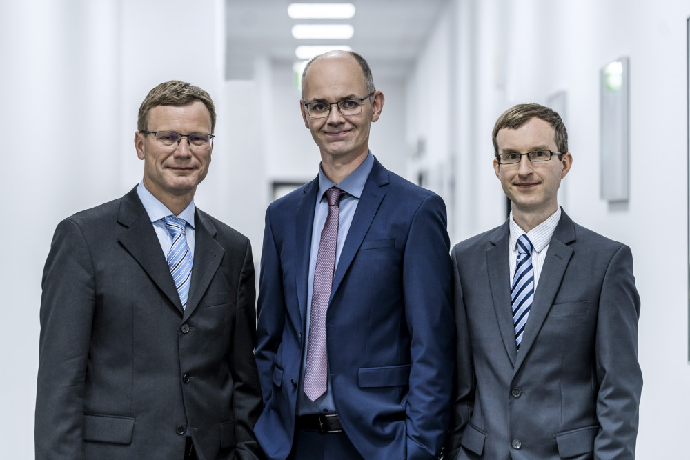
(f.l.t.r.) Dipl.-Phys. Ralf Wolleschensky, Dr. rer. nat. Thomas Kalkbrenner,
Dr. rer. nat. Jörg Siebenmorgen

Dr. Thomas Kalkbrenner, Dr. Jörg Siebenmorgen and Ralf Wolleschensky have a solution: an innovative microscope system. The system opens entirely new perspectives for research in biology, medicine, and pharmacology by linking lattice light sheet microscopy with various innovative optical technologies. This protects sensitive living samples from being damaged by the laser light used during microscopic examination. At the same time, the nominees found a way to make the complex technology easy to use.

This opens the door to a wide range of applications never possible before. Fundamental biological and biomedical research profit from the innovation as well as the search for new approaches to diagnostics and the treatment of diseases. Thomas Kalkbrenner is team lead and Lead Architect R&D Special 3D at Carl Zeiss Microscopy. Jörg Siebenmorgen is Project Manager Development in the Advanced Development division. Ralf Wolleschensky is head of the Advanced Development division.

High-resolution, three-dimensional imaging using fluorescence microscopy has already done a lot for the decoding of the secrets of life. In this method, an organic sample, such as a group of human body cells, is first prepared using a biomarker and then illuminated with laser light of a certain wavelength. The light stimulates the biomarker molecules, causing them to emit fluorescent light. By observing its distribution, a wide range of information about biological processes on a tiny scale can be obtained for example inside a cell.

However, the laser light the biological sample is exposed to can damage it – biological structures and processes undergo a change or are even destroyed. Researchers call this phototoxicity. It can also lead to a false interpretation of images obtained under the microscope. Studies of living systems over a longer period are in many cases not even possible. This narrowly limits the findings that can be obtained by fluorescent imaging. In addition, this limit is all the more drastic the more details are hoped to be observed.
More Details
Resume
Dr. rer. nat. Thomas Kalkbrenner
- April 29, 1971
- Born in Überlingen, Germany
- 1990
- Abitur at Dietrich-Bonhoefer-Gymnasium, Überlingen
- 1991 – 1997
- Studies of physics at the University of Constance, degree: Diplom
- 1998 – 2002
- Research assistant at the University of Constance
- 1998 – 2002
- "Doctorate at the chair of Prof. J. Mlynek under Prof. V. Sandoghdar at the University of Constance"
- 2002 – 2003
- "Scientist at Institute of Physical Chemistry at ETH Zurich, Switzerland"
- 2003 – 2006
- Scientist at FOM Institute AMOLF, Amsterdam, the Netherlands
- 2006 – 2008
- Project Manager Optical Systems at CyBio AG, Jena
- 2008 – 2020
- Project Manager and Technology Specialist in Advanced Development at Carl Zeiss Microscopy GmbH, Jena; main focus: Super-resolution microscopy, camera-based systems, light sheet microscopy
- Since 2021
- Principal in ZEISS Expert Ladder
- Since 2021
- Team Leader and Lead Architect R&D of Special 3D product line
Honors and Awards
- 1998 – 2000
- Carl Zeiss Schott doctoral scholarship
- 2009
- ZEISS Innovation Award, special award for super-resolution microscopy
- 2014
- R&D 100 Award for ZEISS Elyra P1 with 3D super resolution
- 2021
- "ZEISS Innovation Award in the category Leading Edge Technology for the ZEISS Lattice Lightsheet 7"
- 2021
- Nominated as team spokesperson for the Thüringen Innovation Award in the category "Light and Life"
Publications
- Various specialist publications and lectures at conferences and symposiums
Patents
- 396 patent applications in 71 patent families
Dr. Jörg rer. nat. Jörg Siebenmorgen
- July 30, 1979
- Born in Pasewalk, Germany
- 1999
- Abitur at Bargteheide district Gymnasium
- 2000 – 2007
- Studies of physics at the University of Hamburg, degree: Diplom
- 2007 – 2010
- Doctorate at the Institute of Laser Physics under Prof. G. Huber at the University of Hamburg
- 2010
- Scientist at the Institute for Laser Physics at the University of Hamburg
- Since 2011
- Project Manager and Technology Specialist in Advanced Development at Carl Zeiss Microscopy GmbH, Jena; main focus: Light sheet microscopy
- Since 2018
- Senior in ZEISS Expert Ladder
- Since 2019
- R&D Project Manager ZEISS Lattice Lightsheet 7
Honors and Awards
- 2007 – 2010
- Doctoral scholarship from DFG Research Training Group 1355
- 2021
- ZEISS Innovation Award in the category Leading Edge Technology for the ZEISS Lattice Lightsheet 7
Publications
- Various specialist publications and lectures at conferences and symposiums
Patents
- 166 patent applications in 37 patent families
Dipl.-Phys. Ralf Wolleschensky
- October 15, 1972
- Born in Jena, Germany
- 1991
- Abitur at Carl Zeiss special school in Jena
- 1992 – 1998
- Studies of physics at the Friedrich Schiller University Jena, degree: Diplom
- 1995 – 1996
- "Studied abroad at the University of Essex, Colchester, UK
Course: Master of Physics, final year" - 1998 – 2000
- Technology Specialist in microscopy, Carl Zeiss Jena GmbH
"Main focus: optical 3D fluorescence microscopy" - 2000 – 2009
- R&D Project Manager Carl Zeiss Microscopy GmbH, Jena
- Since 2009
- Head of Advanced Development, Carl Zeiss Microscopy GmbH, Jena
- Since 2009
- Senior Principal in ZEISS Expert Ladder
Honors and Awards
- 1996 – 1997
- Scholarship from Carl-Zeiss-Schott Förderstiftung in the Donors' Association for the Promotion of Sciences and Humanities in Germany
- 1998
- Faculty award from Friedrich-Schiller-University in Jena for the best Diplom thesis of the academic year
- 2004
- Nominated for the German Future Prize – German President's Prize for Technology and Innovation
Publications
- Various specialist publications and lectures at conferences and symposiums
Patents
- 687 patent applications in 255 patent families
Contact
Coordination and Press
Beatrice Weinberger
Referentin Presse- und Öffentlichkeitsarbeit
Corporate Brand and Communications
Carl Zeiss AG
Carl-Zeiss-Promenade 10
07745 Jena
Phone: +49 (0) 3641 / 64 23 35
Mobile: +49 (0) 151 / 74 40 09 65
E-Mail: beatrice.weinberger@zeiss.com
Web: www.zeiss.de
Spokesperson
Dr. Thomas Kalkbrenner
Teamleiter R&D Special 3D
Zeiss Research Microscopy Solutions
Carl Zeiss Microscopy GmbH
ZEISS Gruppe
Carl- Zeiss-Promenade 10
07745 Jena
Phone: +49 (0) 3641 / 64 25 34
Mobile: +49 (0) 171 / 38 52 019
E-Mail: thomas.kalkbrenner@zeiss.com
Web: www.zeiss.com/microscopy
A description provided by the institutes and companies regarding their nominated projects
Researching the foundations of life – An innovative microscope for gentle 3D imaging of living cells
Since Robert Koch first observed the bacteria that causes tuberculosis in 1882, the microscope has been at the center of research and development for new therapies and drugs. Indeed, new findings in the life sciences today would be inconceivable without magnifying images of the smallest units of life, such as cells and bacteria. Especially in recent years, enormous progress has been made in cell biology, cancer research, and pharmacology, all of which is based in part on data from fluorescence microscopes. Thus, to advance medical progress as a whole, one must first succeed in improving microscopy. With this in mind, Dr. Thomas Kalkbrenner, Dr. Jörg Siebenmorgen, and Ralf Wolleschensky, together with their team, have succeeded in revolutionizing fluorescence microscopy with the development of the ZEISS Lattice Lightsheet 7.
Living cells are harmed by observation
When it comes to studying living cells with fluorescence microscopes, illumination poses a particular challenge to scientists: The intensities of the laser radiation used exceed those of the sun by a factor of 1000 – the light source to which life on our planet has adapted. Thus, it is not surprising that intense illumination like this can cause permanent damage to living cells and even kill them.
A paradigm shift is needed to overcome this challenge. The ZEISS Lattice Lightsheet 7 features several innovations that push the boundaries of fluorescence microscopy to open up new possibilities for research and development in the life sciences.
Lattice light sheets: A unique illumination concept tames laser beams
A solution referred to as light sheet microscopy allows for a significant reduction in damage caused by radiation (photodamage): Unlike all other microscopes, the laser radiation – in the form of a light sheet – is introduced only into the area that is in the focus of the objective. The method significantly reduces the amount of radiation the organism under inspection is exposed to. Developmental biologists, who can use the innovation to study larger model organisms such as fruit flies over long periods of time, stand to benefit above all.
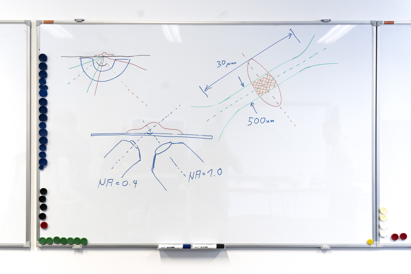
However, the laws of optics prevent the application of this technology in cell biology: Focusing traditional beams tighter to achieve thinner light sheets needed for subcellular resolution also shortens the beams – there’s no light sheet anymore.
So, as a first challenge, non-traditional beam shapes had to be developed that allow light sheets that are both very thin and long at the same time. Professor Eric Betzig developed a solution to this problem with so-called lattice light sheets. They permit, for the first time, biological processes at the subcellular level to be observed over a period of hours or even days.
But the generation and application of these special light sheets is both highly complex and very time-consuming, meaning that those systems have to be operated by specialists. The team has further improved this complex beam shaping process, automating it to the point where users can select optimal light sheets for their particular application with the click of a mouse.
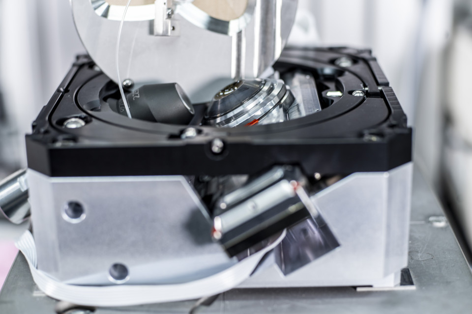
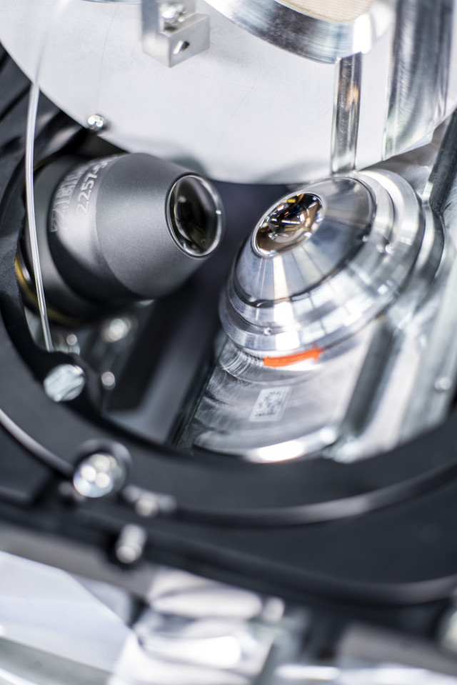
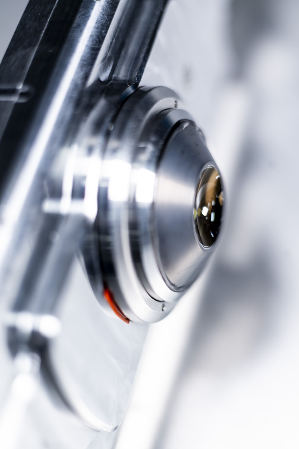
Looking through the glass at an angle
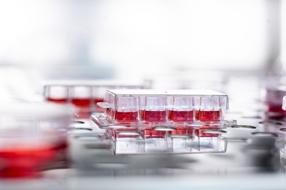
This improvement notwithstanding, the widespread use of this fascinating technology in research and drug development remains out of reach: Cells grow on coverslips in culture vessels such as Petri dishes and multiwell plates, which, due to the special arrangement of the objectives, cannot be used in a traditional light sheet microscope. One would have to look from below at an angle through the cover glass – an impossible task for a microscope objective, because the distortions that occur prevent any imaging. So unique microscope optics were developed that corrects these image errors with the help of adaptive freeform elements, for any sample containers, even those of varying thickness. Manufacturing technologies from ZEISS semiconductor optics are used to create these special optical elements.
This optical core now allows the widespread use of the revolutionary light sheet technology without any limitations in sample preparation. In particular, the multiwell plate formats that are so important for drug development (high-content screening) are made accessible.
Gentle on living cells, fast, and easy to operate
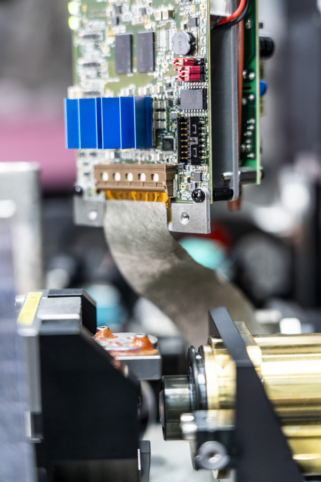
All this has been developed into an easy-to-use, compact system with high potential for automation. The software offers special coordinated workflows to quickly convert the raw data into a coordinate system familiar to researchers for further processing. Unlike conventional microscope systems, the system always generates 3D data, and does so with near-isotropic 3D resolution and at multiple volumes per second. This results in data rates of up to 1.4 gigabytes per second; the data are recorded, stored, and processed with an optimized computer and software architecture.
The nominees and their team have thus developed a microscope system that not only surpasses the optical properties of lattice light sheet laboratory systems but can also be easily operated by any scientist.
New science in practice
With its revolutionary technology, ZEISS Lattice Lightsheet 7 enables biomedical researchers, for the first time in practice, to observe living cells in 3D for hours or even days. This lets them investigate for example how living cells react to certain active substances, or what happens when viruses or bacteria enter cells. The system impresses not only with its gentle treatment of the sample, but also with its high temporal resolution. Even processes that take less than a second can now be made visible in 3D.
Dr. Eric Rentchler from the University of Michigan (USA) was among the first to test ZEISS Lattice Lightsheet 7. "They were blown away by what they were able to see," describes Dr. Rentchler his team's first impressions of the microscope. "They were noticing some phenomena that they’re still trying to explain – something new that they've never observed before." This means ZEISS Lattice Lightsheet 7 is the only commercially available system that makes the unrivaled sample protection of lattice light sheets accessible to any researcher, even those without special prior knowledge. With ZEISS Lattice Lightsheet 7, ZEISS is providing researchers in biology, medicine, and pharmaceutics with a valuable tool for unraveling both the secret mechanisms of life and its pathologies, and for harnessing this research for the benefit of all.
Outlook
Since the market launch at the end of 2020, the platform has already been updated. The focus was a further increase in recording speed, improvements in data handling, and even higher levels of automation. The next step is to combine ZEISS Lattice Lightsheet 7 with super-resolution methods (2014 Noble Prize). The system concept fits perfectly here, and will then enable gentle imaging even below the resolution limit of optical imaging systems.
Just as it took the observation of the bacteria responsible for tuberculosis in 1882 to develop effective treatments for the disease, the use of ZEISS Lattice Lightsheet 7 in research marks a breakthrough for new treatment approaches in the future. Biological processes such as signal transduction, receptor interactions, intracellular transport mechanisms, or even the infection of a cell by bacteria or viruses can be observed over longer periods of time, which will lead to completely new insights and therapies. Additionally, through the use of multiwell plates, the system's unique optical core enables gentle 3D high-content screening, a method crucial in drug development. The upshot is that so-called organoids are literally coming into focus: As small, three-dimensional model organs made of human tissue, they possess great potential, especially in cancer research, and pose an alternative to drug validation in traditional animal experiments. But, at the same time, they are also very sensitive to light – and thus the perfect challenge for ZEISS Lattice Lightsheet 7.
"They were blown away by what they were able to see. They were noticing some phenomena that they're still trying to explain – something new that they've never observed before."
Dr. Eric Rentschler, University of Michigan
About ZEISS
ZEISS is an internationally leading technology enterprise operating in the fields of optics and optoelectronics. In the previous fiscal year, the ZEISS Group generated annual revenue totaling 7.5 billion euros in its four segments Semiconductor Manufacturing Technology, Industrial Quality & Research, Medical Technology and Consumer Markets (status: 30 September 2021).
For its customers, ZEISS develops, produces and distributes highly innovative solutions for industrial metrology and quality assurance, microscopy solutions for the life sciences and materials research, and medical technology solutions for diagnostics and treatment in ophthalmology and microsurgery. The name ZEISS is also synonymous with the world's leading lithography optics, which are used by the chip industry to manufacture semiconductor components. There is global demand for trendsetting ZEISS brand products such as eyeglass lenses, camera lenses and binoculars.
With a portfolio aligned with future growth areas like digitalization, healthcare and Smart Production and a strong brand, ZEISS is shaping the future of technology and constantly advancing the world of optics and related fields with its solutions. The company's significant, sustainable investments in research and development lay the foundation for the success and continued expansion of ZEISS' technology and market leadership. ZEISS invests 13 percent of its revenue in research and development – this high level of expenditure has a long tradition at ZEISS and is also an investment in the future.
With around 37,000 employees, ZEISS is active globally in almost 50 countries with around 30 production sites, 60 sales and service companies and 27 research and development facilities (status: 31 March 2022). Founded in 1846 in Jena, the company is headquartered in Oberkochen, Germany. The Carl Zeiss Foundation, one of the largest foundations in Germany committed to the promotion of science, is the sole owner of the holding company, Carl Zeiss AG.
Carl Zeiss Microscopy GmbH belongs to the ZEISS segment Industrial Quality & Research.
Further information at www.zeiss.com
The right to nominate outstanding achievements for the Deutscher Zukunftspreis is incumbent on leading German institutions in science and industry as well as foundations. The project " Researching the foundations of life – An innovative microscope for gentle 3D imaging of living cells” was submitted by Deutsches Patent und Markenamt.
Federal President Frank Walter Steinmeier will present the Deutscher Zukunftspreis to one of the three nominated teams on October 26, 2022.