 Gebärdensprache
 Leichte Sprache

Nominee 2021
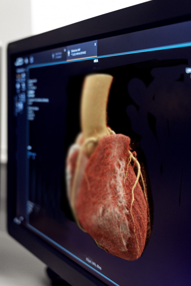
Photon-counting computed tomography scanner

(f.l.t.r.) Dr. rer. biol. hum. Stefan Ulzheimer, Prof. Dr. rer. nat. Thomas Flohr,
Dr. rer. nat. Björn Kreisler

Professor Thomas Flohr, Dr. Björn Kreisler and Dr. Stefan Ulzheimer have opened the door to a new application. The three nominees have developed technical components and a number of innovative techniques to detect the X-ray light used in computer tomography. They also re-structured the CT system's digital architecture.

This makes it possible to obtain images of the human body in a detail previously impossible. The innovative detector principle is based on the counting and analysis of individual X-ray photons using crystalline materials. This produces a spatial resolution previously unattainable in computer tomography images and also provides much valuable additional information.


More Details
Resume
Prof. Dr. rer. nat. Thomas Georg Flohr
- 12.01.1960
- Born in Neustadt / Aisch, Germany
- 1970 - 1979
- Qualification: High-school diploma, Friedrich-Alexander-Gymnasium, Neustadt / Aisch, Germany
- 1979 - 1985
- Study of physics, qualification: Diploma, Friedrich-Alexander-University, Erlangen-Nuremberg Erlangen, Germany
- 1985 - 1989
- Doctoral candidate in physics, Friedrich-Alexander-University, Erlangen-Nuremberg Erlangen, Germany
- 1989
- Qualification: Doctor of natural sciences (Dr. rer. nat), Friedrich-Alexander-University Erlangen-Nuremberg, Erlangen, Germany
- 1989 - 2000
- Research position in the computed tomography section, Siemens, UB Med, Erlangen/Forchheim, Germany
- 2000 - 2003
- Head of CT physics department, computed tomography section, Siemens, UB Med, Forchheim, Germany
- Since 2004
- Head of the CT physics, application predevelopment, and global clinical collaborations department, Siemens Healthcare, now Siemens Healthineers, Forchheim, Germany
- 2006
- Habilitation in "Medical Physics", Eberhard-Karls-University Tuebingen, Tuebingen, Germany
- 2007 - 2011
- Qualified Professor, Eberhard-Karls-University Tuebingen, Tuebingen, Germany
- Since 2011
- Adjunct professor for medical physics, Eberhard-Karls-University Tuebingen, Tuebingen, Germany
Further activities
- Since 2005
- Founding member of the Society of Cardiovascular Computed Tomography
- Since 2006
- Member of the Program Committee of the Physics of Medical Imaging Conference of the SPIE
- Since 2007
- Member of the Scientific Committee of the Fully 3D Conference: International Meeting on Fully Three-Dimensional Image Reconstruction in Radiology and Nuclear Medicine
- 2016 - 2017
- Conference Chair of the Physics of Medical Imaging Conference of the SPIE
Other
- 230 peer-reviewed publications in scientific journals, including 46 as the first or last author
Publisher / author of five books
Ad hoc reviewer for several scientific journals, including Investigative Radiology, European Radiology, Medical Physics
Awards (a selection)
- 1998
- "Inventor of the Year" of Siemens AG for work on the 4-slice CT scanner "SOMATOM Volume Zoom"
- 2002
- Finalist of the German Future Prize of the German Federal President, for "Looking into the heart without a catheter – cardio CT” (Bernd Ohnesorge, Thomas Flohr, Richard Hausmann)
- 2003
- Winner of the Siemens "top+ award" in the category "Innovation" for the 16-slice CT scanner "SOMATOM Sensation 16"
- 2007
- Appointment as "Siemens Top Innovator"
- 2009
- Winner of the Siemens "top+ award" in the category "Innovation" for the dual-source CT scanner "SOMATOM Flash"
- 2015
- Finalist of the German Innovation Prize for the Dual Source CT Scanner "SOMATOM Force"
- 2021
- Award of an honorary doctorate from the Faculty of Medicine of the University of Zurich
Award-winning papers
- The paper "Flohr TG et al, First performance evaluation of a dual-source CT (DSCT) system. Eur Radiol. 2006" was chosen as one of 10 papers for presentation at the "European Radiology 25th Anniversary Session" at the ECR 2017. It is among the 100 most cited works in radiology journals in the years 1945-2012 (Yoon DY et al. Citation Classics in Radiology Journals: The 100 Top-Cited Articles, 1945-2012. American Journal of Roentgenology. 2013;201: 471-481
The paper "Ohnesorge B, Flohr T, et al. Cardiac imaging by means of electrocardiographically gated multisection spiral CT: initial experience. Radiology. 2000" was included in the series "Radiology Golden Oldies" (Radiology. 2015;276(3))
DThe paper "Flohr TG, et al. Dual-source spiral CT with pitch up to 3.2 and 75 ms temporal resolution: image reconstruction and assessment of image quality. Med Phys. 2009" was chosen for the edition "Significant Advances in CT – an inaugural Virtual Issue of Medical Physics in honor of the 40th anniversary of Cormack and Hounsfield’s 1979 Nobel Prize for the development of the CT scanner" of the journal Medical Physics.
Dr. rer. nat. Björn Kreisler
- 10.02.1980
- Born in Frankfurt am Main, Germany
- 1990 - 1999
- Qualification: High-school diploma, Emil-von-Behring Gymnasium, Spardorf, Germany
- 1999 - 2000
- Civilian service, Erlangen, Germany
- 2000 - 2006
- Studies in physics, focus "Physics in medicine", Friedrich-Alexander-University, Erlangen-Nuremberg / University of York, Erlangen, Germany / York, United Kingdom
- 2006
- Diploma in physics, thesis: "Influenced signals in pixelated semiconductor X-ray detectors”, Friedrich-Alexander-University Erlangen-Nuremberg, Erlangen, Germany
- 2006 - 2010
- Research position in the Medical Physics Group at the Erlangen Center for Astroparticle Physics (ECAP) in the Institute for Physics, Friedrich-Alexander-University Erlangen-Nuremberg, Erlangen, Germany
- 2010
- Doctorate at the Institute for Physics: "Simulation of Medical Irradiation and X-Ray Detector Signals", Friedrich-Alexander-University Erlangen-Nuremberg, Erlangen, Germany
- Since 2010
- Detector physicist for CT detectors with the focus on counting detectors, Siemens Healthcare, now Siemens Healthineers, Forchheim, Germany
- 2019
- Appointment as senior key expert for detectors, Siemens Healthineers, Forchheim, Germany
Study grants
- 2002 - 2003
- Erasmus grant for study year at York, United Kingdom
- 2006 - 2010
- Doctoral studies grant of the International Max-Planck Research School (IMPRS) for Optics and Imaging
Dr. rer. biol. hum. Stefan Ulzheimer
- 12.04.1971
- Born in Nuremberg, Germany
- 1981 - 1990
- Gymnasium, Martin-Behaim-Gymnasium, Nuremberg, Germany
- 1990
- Qualification: High-school diploma, Martin-Behaim-Gymnasium, Nuremberg, Germany
- 1990 - 1991
- Basic military service, Regensburg, Germany
- 1991 - 1994
- Foundation studies in physics, Friedrich-Alexander-University Erlangen-Nuremberg, Erlangen, Germany
- 1993 - 1994
- Erasmus student, Imperial College, London, United Kingdom
- 1994
- Intermediate examination in physics, Friedrich-Alexander-University Erlangen-Nuremberg, Erlangen, Germany
- 1994 - 1998
- Advanced studies in physics, Friedrich-Alexander-University Erlangen-Nuremberg, Erlangen, Germany
- 1998
- Qualification: Diploma in physics, Friedrich-Alexander-University Erlangen-Nuremberg, Erlangen, Germany
- 1998 - 2001
- Doctoral candidate in human biology at the Faculty of Medicine, Friedrich-Alexander-University Erlangen-Nuremberg, Erlangen, Germany
- 2000
- Doctor of human biology (Dr. rer. biol. hum.), Friedrich-Alexander-University Erlangen-Nuremberg, Erlangen, Germany
- 2001 - 2004
- General manager, Verfahren und Apparate der Medizinischen Physik GmbH, Moehrendorf, Germany
- 2004 - 2009
- Collaboration manager for computed tomography, Siemens Healthcare, Forchheim, Germany
- 2010 - 2014
- Head of Global Scientific Marketing for Computed Tomography, Siemens Healthcare, Forchheim, Germany
- Since 2014
- Program Director for counting technology, Siemens Healthcare, now Siemens Healthineers, Forchheim, Germany
Awards
- 1999/2000
- Magna cum laude prize of the ECR (European Congress of Radiology): "Improvements of cardiac CT using ECG oriented image reconstruction in subsecond spiral multirow scanning"
- 2001
- Winner of the Hochschulgründerpreis (prize for university spin-offs) in the Bavarian Business Plan Competition: "TomoScope – A micro-CT system for in-vivo small animal imaging"
Contact
Press
Ulrich Künzel
Communications, Media Relations
Siemens Healthcare GmbH
Karlheinz-Kaske-Straße 5
91052 Erlangen
Mobile: +49 (0) 162 / 24 33 492
E-Mail: ulrich.kuenzel@siemens-healthineers.com
www.siemens-healthineers.com
Spokesperson
Prof. Dr. Thomas Flohr
Siemens Healthcare GmbH
Siemensstraße 3
91301 Forchheim
Mobile: +49 (0) 173 / 25 62 351
E-Mail: thomas.flohr@siemens-healthineers.com
A description provided by the institutes and companies regarding their nominated projects
Photon-counting computed tomography scanner – A revolutionary way of looking inside the human body
Computed tomography (CT) is an indispensable tool of modern medicine for early and fast detection and diagnosis of diseases. It is the backbone of radiological diagnosis. Yet, not all diagnostic questions can be answered using CT imaging. For some diseases, a CT examination will not produce a conclusive diagnosis from which necessary treatment steps can be derived. In such cases, further examinations with other procedures will be required to diagnose the disease reliably. There are also various reasons why certain patient groups are not examined with fast and straightforward CT imaging, which can delay reporting. These reasons include reticence regarding X-ray dose in the case of children and young adults and the commonly used iodinated contrast media in the case of kidney disease patients.
Conventional CT imaging is a technically mature procedure, so further efforts to overcome these limitations in order to achieve earlier, more conclusive diagnosis of larger patient groups is not to be expected. Nominees Professor Thomas Flohr, Dr. Bjoern Kreisler, and Dr. Stefan Ulzheimer have overcome this obstacle. Together with their colleagues at Siemens Healthineers, they developed an innovative CT concept, tested it with clinical prototypes, and developed it for series production. The concept is based on completely new detector technology and thorough redevelopment of all the system components. Photon-counting computed tomography paves the way for the previously unattainable combination of strong image sharpness and extremely short image acquisition times with improved image contrast and more diagnostically useful image contents. At the same time, the X-ray and contrast agent dose can be reduced. This is not merely a new, improved generation of CT scanners – CT imaging has in effect been reinvented.
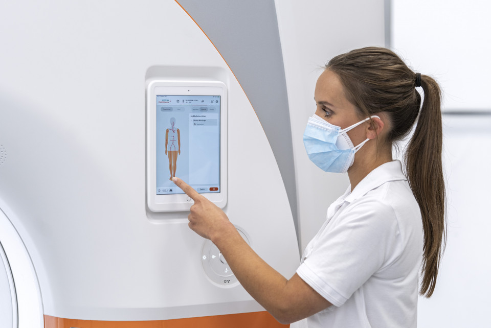
In a CT scanner, an X-ray tube and a detector system rotate around the patient. During this rotating movement, the detector registers the X-rays attenuated by the body and converts them into an electrical signal, which is then digitized. From this data, a computer calculates images that show the inside of the body and its organs in three dimensions and without overlap. Conventional detectors operate in two stages: The X-rays are converted to visible light in a scintillator, which then generates an electrical signal. The information about the energy of the X-rays is lost, which results in reduced image contrast and unclear image content. It is also not possible to reduce the size of the individual detector elements meaning that image sharpness cannot be significantly increased.
Significantly wider range of applications for computed tomography
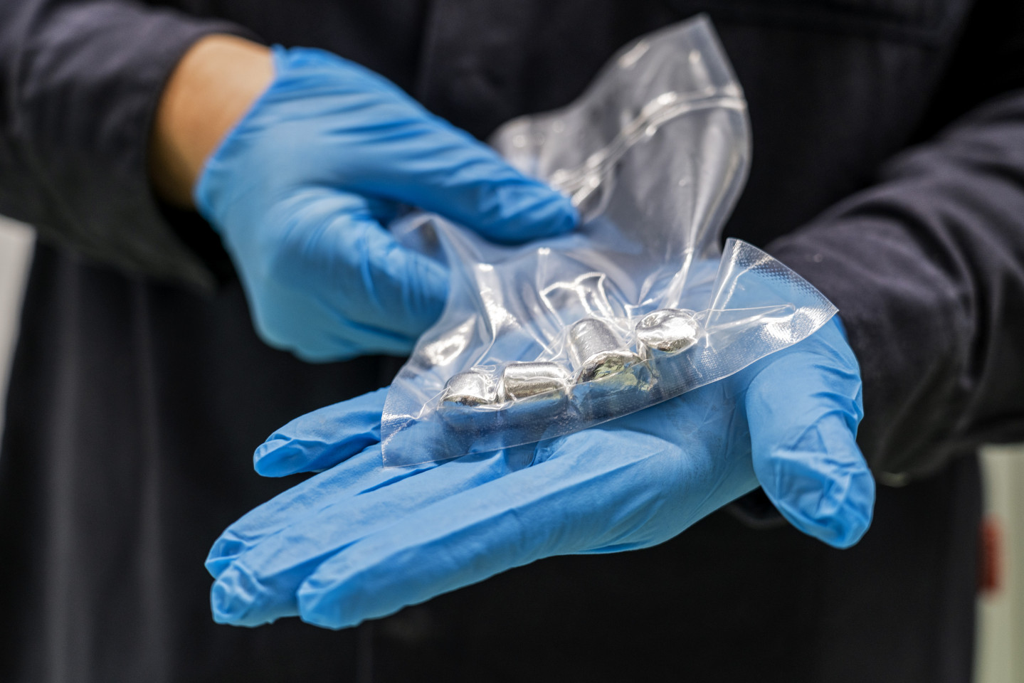
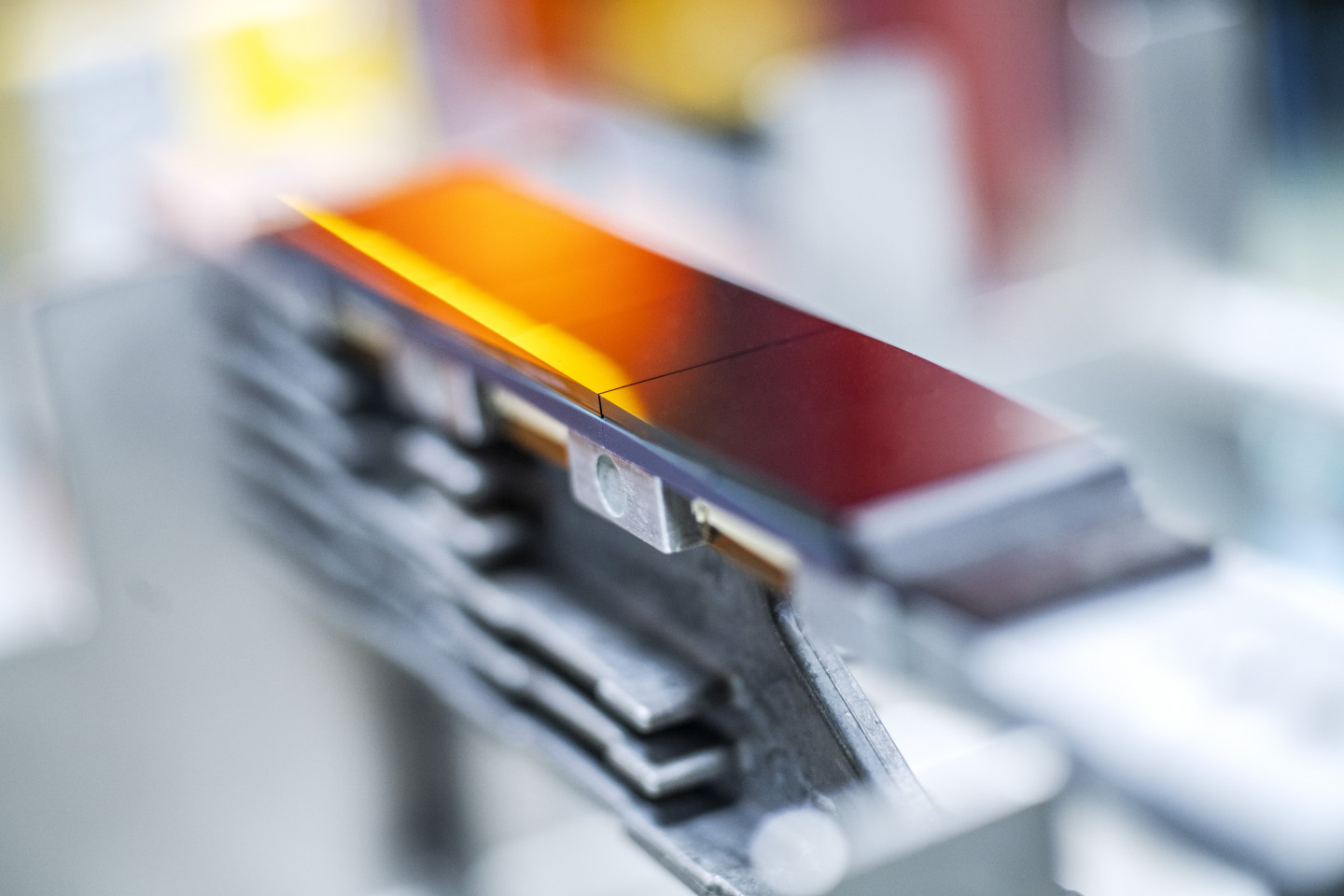
As early as 2001, the Siemens healthcare division embarked on the development of new detector concepts in cooperation with partner organizations from research and industry. Cadmium telluride (CdTe) was soon identified as a very promising material. The "photon-counting detector" converts X-rays in a CdTe crystal into electrical pulses, whose magnitude is proportional to the energy of the X-ray. The energy information is not lost when the detector is read out – this increases image contrasts, the images contain more information and are clearer, making diagnosis easier. The fine structures of the detectors result in a considerable increase in image sharpness, and the X-ray and contrast medium dose can be substantially reduced with the new detection concept. As a result, photon-counting CT has extended the range of use to patient groups for whom CT imaging was previously considered unsuitable due to the X-ray or contrast agent dose. When the new technology is implemented in a Dual Source CT – that is, in a system with two X-ray tubes and two detectors – extremely short image acquisition times and fast acquisitions are achieved. As a result, the examination of moving organs, such as the lung and heart, is possible with previously unknown accuracy.
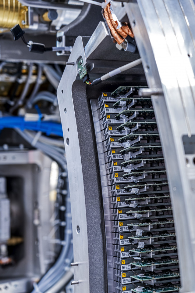
These improvements would not have been possible with conventional CT technology. The CdTe detector material available in 2001 was, however, far from meeting the high-quality requirements of medical CT. During the course of nearly 20 years of development work, Siemens Healthineers and its partners took the technology step by step to series maturity with numerous improvements and laid the foundations for series detector manufacturing. With completely redeveloped readout electronics connected to the CdTe crystals across their entire surface, any number of individual crystals can be juxtaposed to form larger detectors. In parallel to this work, the three nominees and their team overhauled and optimized the entire CT system concept, its hardware and software, to make the best possible use of the advantages of the new detector. In order to handle the significantly higher data volume and process data containing much more information, new approaches to data transfer and algorithms were developed. Three-dimensional images are now calculated in a matter of seconds on much more powerful computers being used for the first time in medical CT systems.
40 percent lower X-ray and contrast medium dose, twice as sharp images, and more refined tissue characterization
Since 2014, prototype CT scanners with these new photon-counting detectors have been installed in clinical settings to examine patients. Considerable knowledge about their potential in clinical use has been collected as a result. The clinical institutions confirmed the predicted reduction of up to 40 percent in X-ray and contrast medium dose on the patients they examined. They achieved significantly stronger image contrasts, for example, in examinations of the brain, and improved tissue characterization. With almost twice the image sharpness, new opportunities in bone and vessel imaging result, leading to more precise and patient-friendly intervention planning, for example, in partial nephrectomy in the treatment of kidney tumors. Lung and heart examinations also benefit from the combination of strong image sharpness, short acquisition times, and more refined tissue characterization. COVID-19 follow-up checks can also be significantly improved in this way. CT examinations of the coronary vessels can now be performed on significantly more patients, even those with severe calcification or stents. Previously, these patients usually had to undergo invasive diagnostic cardiac catheterization. With the new CT technology, diagnostic images of the coronary vessels with reliable visualization of stenoses can be acquired in these cases, too – without the risks of an invasive examination. In the case of polytraumas, the whole body can be examined with the highest precision in under three seconds.
In trials of the new CT technology, the nominees also recognized opportunities for further improving imaging with new contrast agents which could be developed by adapting them to the properties of the photon-counting detector. This was taken up by Bayer AG, who as part of a partnership with Siemens Healthineers is already researching new CT contrast agents.
Overall, a large number of cooperation partners has been recruited over the 20-year history of this unique research and development project. New core competencies have been established and transferred to the project. Company locations have been expanded, new employees recruited, a new production center for CdTe crystals built in Forchheim, and 30 leading clinical research partners have been involved. These include, for example, the university hospitals in Augsburg, Tübingen, Freiburg, and Hanover, as well as University Hospital Zurich and Erasmus Medical Center Rotterdam.
In future, every computed tomography scanner will be a photon-counting CT
The huge potential of this innovative CT concept offers great advantages for doctors and patients alike. Patients who previously could not benefit from this noninvasive, fast examination method can now be examined with CT imaging. Moreover, the technology offers opportunities for more precise, earlier diagnoses with fewer time-consuming and costly additional examinations, better therapy decisions, and an earlier therapy start. Another possibility is the more extensive utilization of the large volume of information extracted with photon-counting CT using artificial intelligence, both to improve cardiovascular and oncological differential diagnosis, and to plan and follow targeted therapies.
Since Siemens Healthineers is confident of the new technology's success, it will be installed in every computed tomography scanner manufactured by the company in the medium term and long term. Independent market forecasts expect an increase in global annual sales of CT systems from 6.43 billion U.S. dollars in 2020 to 9.64 billion U.S. dollars in the year 2027, which could be replaced with photon-counting CT systems. Due to its innovation and market lead of several years, Siemens Healthineers with its production and development location Forchheim will have a disproportionately larger share of this market. This development will also further accelerate contrast media research and contribute to shaping new research areas in process engineering and intelligent processing of large data volumes.
Siemens Healthineers AG (listed in Frankfurt, Germany: SHL) is shaping the future of healthcare. As a leading medical technology company headquartered in Erlangen, Germany, Siemens Healthineers enables healthcare providers worldwide through its regional companies to increase value by empowering them on their journey towards expanding precision medicine, transforming care delivery, improving the patient experience, and digitalizing healthcare. Siemens Healthineers is continuously developing its product and service portfolio, with AI-supported applications and digital offerings that play an increasingly important role in the next generation of medical technology. These new applications will enhance the company’s foundation in in-vitro diagnostics, image-guided therapy, in-vivo diagnostics, and innovative cancer care. Siemens Healthineers also provides a range of services and solutions to enhance healthcare providers’ ability to provide high-quality, efficient care to patients. In fiscal 2020, which ended on September 30, 2020, Siemens Healthineers generated revenue of €14.5 billion and adjusted EBIT of €2.2 billion. Following the acquisition of Varian Medical Systems, Inc. the company has approximately 66,000 employees worldwide. Further information is available at www.siemens-healthineers.com.