Gebärdensprache
 Leichte Sprache


Nominee 2004
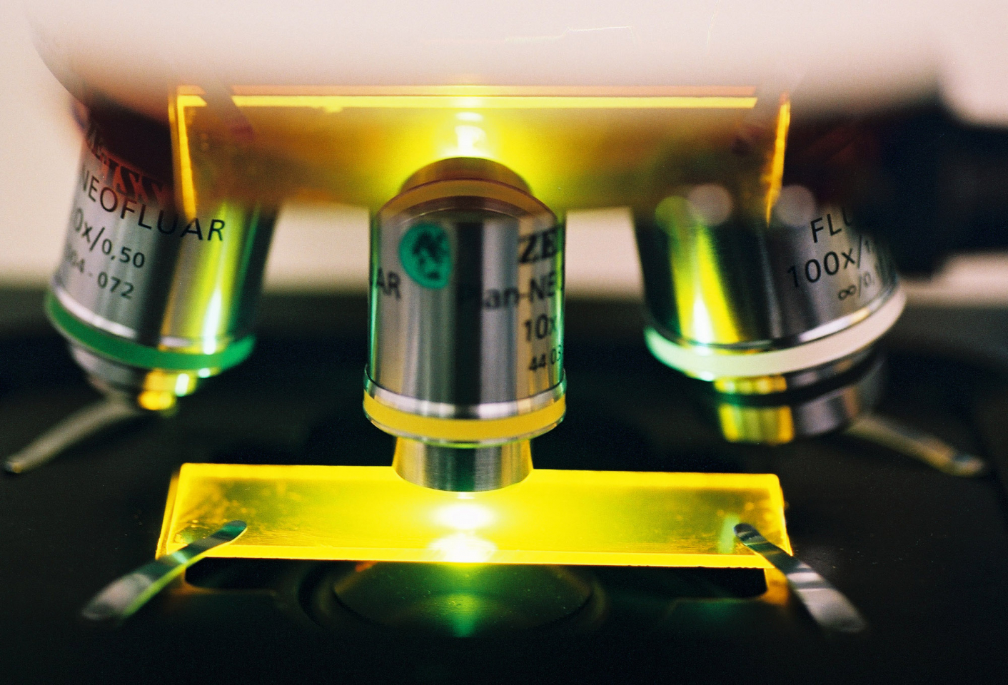
Laser Scanning Mikroskop
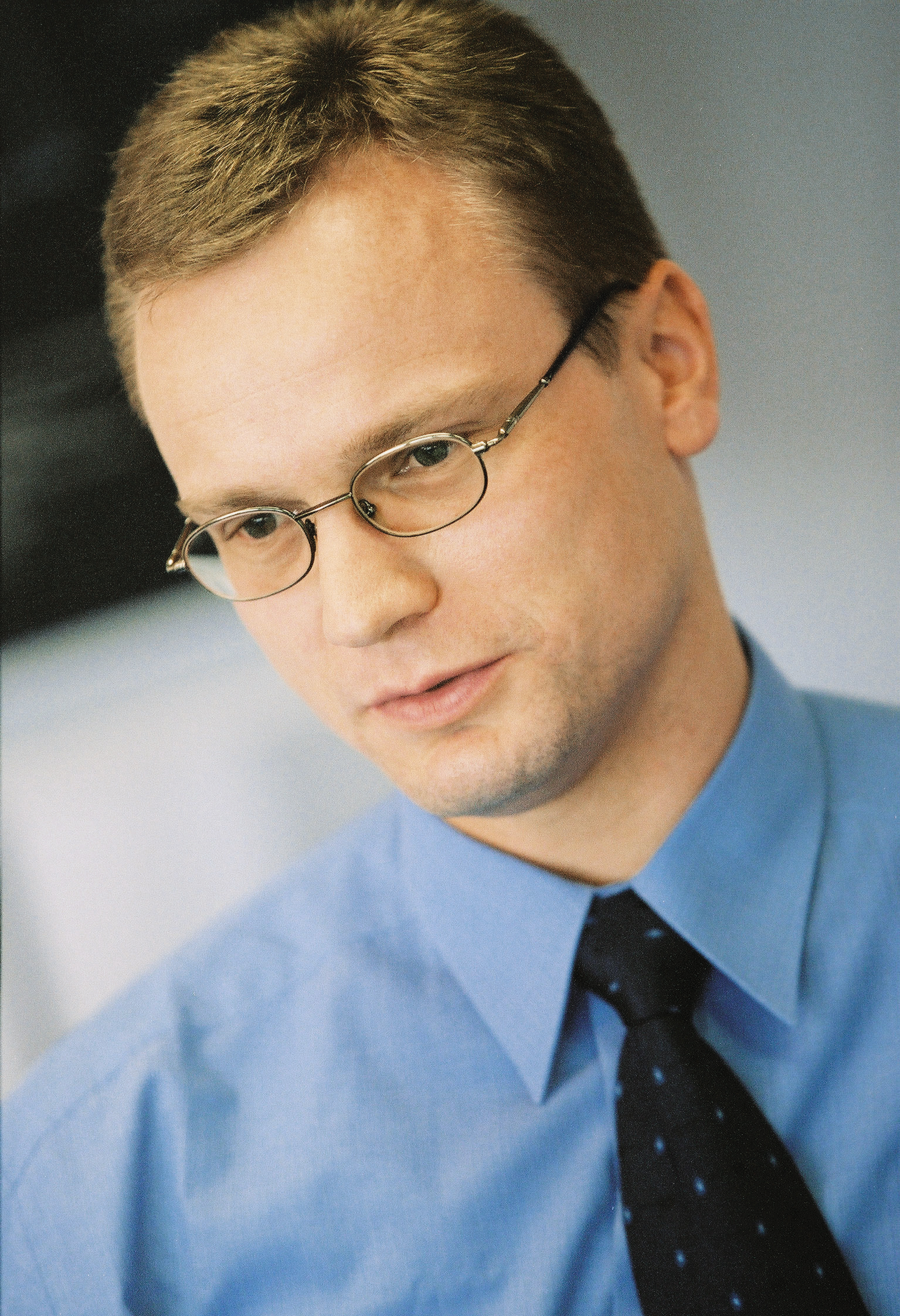
 Dr. rer. nat. Bernhard Zimmermann, Dr. rer. nat. Dipl.-Phys. Ulrich Simon, Dipl.-Phys. Ralf Wolleschensky")
(f.l.t.r.) Dr. rer. nat. Bernhard Zimmermann, Dr. rer. nat. Dipl.-Phys. Ulrich Simon, Dipl.-Phys. Ralf Wolleschensky

Ulrich Simon, Ralf Wolleschensky and Bernhard Zimmermann succeeded in reaching this goal with an innovative detection process. All three of the nominated researchers work for Carl Zeiss Jena GmbH: Ulrich Simon heads the division Microscopy as Executive Vice President and General Manager, Ralf Wolleschensky is development project manager for Advanced Imaging Microscopy, and Bernhard Zimmermann heads product management for the division.
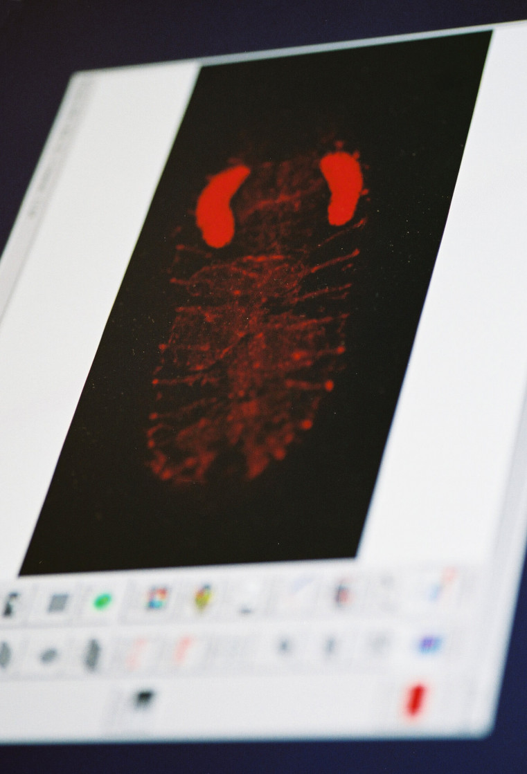
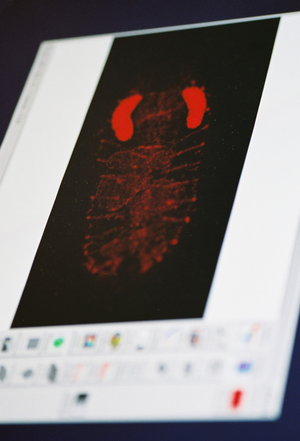
A laser lets marked proteins glow
Biomedical research uses laser microscopy to identify cellular structures. This technology makes structures and processes of movement in cells visible with high spatial accuracy. Functions and dysfunctions can be recognized on the images – and can be used to detect defective growth or, for example, tumors.


more details
Resumes
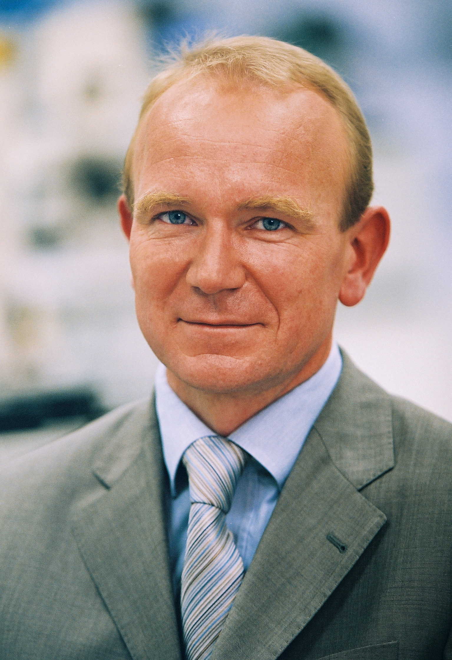
Dr. rer. nat. Dipl.-Phys. Ulrich Simon
- 15.10.1963
- geboren in Bonn
- 1982 – 1989
- Studium der Physik an der Rheinischen Friedrich-Wilhelm Universität Bonn
- 1983
- Praktikum in Elektronik am Forschungszentrum für Angewandte Naturwissenschaften E. V. (FGAN), Wachtberg-Werthoven
- 1985
- Praktikum in Laserphysik am Institut für Angewandte Physik (IAP), Universität Bonn
- 1989 – 1991
- Wissenschaftlicher Mitarbeiter, Universität Bonn
- 1991
- Promotion in Physik, Universität Bonn
- 1992 – 1993
- Robert A. Welch Postdoctoral Fellow, William Marsh Rice University, Houston, Texas, USA
- 1993 – 1994
- Feodor Lynen Fellow der Alexander-von-Humboldt-Stiftung, William Marsh Rice University, Houston, Texas, USA
- 1993
- Forschungsaufenthalt im Naval Research Laboratory (NRL), Washington, D.C., USA
- 1993, 1994
- Forschungsaufenthalte am National Institute of Standards and Technology (NIST), Boulder, Colorado, USA
- 1994 – 1995
- Entwicklungsingenieur für die Laser Scanning Mikroskope im Unternehmensbereich Mikroskopie, Carl Zeiss Jena GmbH
- 1995 – 1996
- Projektleiter für das Laser Scanning Mikroskop LSM 510 META, Carl Zeiss Jena GmbH
- 1996 – 1998
- Entwicklungsleiter Scanning Mikroskopie, Carl Zeiss Jena GmbH
- 1998 – 1999
- Produktgruppenleiter Scanning Mikroskopie, Carl Zeiss Jena GmbH
- 1999 - 2002
- Geschäftsbereichsleiter (Vice President and General Manager) des Geschäftsbereiches Advanced Imaging Microscopy, Carl Zeiss Jena GmbH
- seit 2002
- Unternehmensbereichsleiter (Executive Vice President and General Manager) des Unternehmensbereiches Mikroskopie, Mitglied der Gruppenleitung der Carl Zeiss AG, Jena
Dipl.-Phys. Ralf Wolleschensky
- 15.10.1972
- geboren in Jena
- 1992 – 1998
- Studium der Physik an der Friedrich-Schiller-Universität, Jena
- 1995 – 1996
- Auslandsstudium der Physik an der University of Essex, Colchester, UK
- 1997
- Förderstipendium der Carl-Zeiss-Schott-Förderstiftung im Stifterverband für die Deutsche Wissenschaft
- 1998
- Diplom an der Friedrich-Schiller-Universität, Jena
- 1998 – 2000
- Wissenschaftlicher Mitarbeiter im Unternehmensbereich Mikroskopie, Carl Zeiss Jena GmbH
- seit 2000
- Entwicklungsprojektleiter im Geschäftsbereich Advanced Imaging Microscopy, Carl Zeiss Jena GmbH
Ehrungen:
- 1999
- Fakultätspreis der Friedrich-Schiller-Universität Jena für die beste Diplomarbeit des Studienjahres

Dr. rer. nat. Bernhard Zimmermann
- 18.08.1962
- geboren in München
- 1981 – 1987
- Studium der Biologie (Diplom) an der Universität Regensburg
- 1987 – 1989
- Ziviler Ersatzdienst am Institut für Zell-Pathologie der Gesellschaft für Strahlen- und Umweltforschung, München
- 1989 – 1992
- Promotion in Biologie, Universität Regensburg
- 1992 – 1993
- Post-Graduierten Studien am Institut für Biologie I, Universität Regensburg
- 1993 – 1994
- Ausbildungsstipendium der Deutschen Forschungsgemeinschaft am Department of Molecular Physiology and Biological Physics, School of Medicine, University of Virginia, Charlottesville, USA
- 1994 – 1995
- Research Scientist am Department of Molecular Physiology and Biological Physics, School of Medicine, University of Virginia, Charlottesville, USA
- 1995 – 2001
- Wissenschaftlicher Angestellter/Assistent am Institut für Biochemie und Biologie, Lehrstuhl für Zoophysiologie der Universität Potsdam
- 2001 – 2003
- Produktmanager für konfokale Laser Scanning Mikroskope im Geschäftsbereich Advanced Imaging Microscopy, Carl Zeiss Jena GmbH
- seit 2003
- Leiter Produktmanagment im Geschäftsbereich Advanced Imaging Microscopy, Carl Zeiss Jena GmbH
Contact
Spokesperson
Dr. rer. nat. Dipl.-Phys. Ulrich Simon
Carl Zeiss Jena GmbH
Leiter des Unternehmensbereiches Mikroskopie
Carl-Zeiss-Promenade 10
07745 Jena
Tel.: +49 (0) 3641 / 64 32 22
Fax: +49 (0) 3641 / 64 33 47
E-Mail: u.simon@zeiss.de
Press
Gudrun Vogel
Carl Zeiss Jena GmbH
Kommunikation
Carl-Zeiss-Promenade 10
07745 Jena
Tel.: +49 (0) 3641 / 64 27 70
Fax: +49 (0) 3641 / 64 29 41
E-Mail: g.vogel@zeiss.de
A description provided by the institutes and companies regarding their nominated projects
Laser microscopes make molecular structures in living cells visible to the eye – an important tool when tracking down cancerous tumors. The difficulty: it is often impossible to differentiate between the different types of molecules.
How can laser microscopes help us see more?
Ulrich Simon, Ralf Wolleschensky and Bernhard Zimmermann succeeded in reaching this goal with an innovative detection process. All three of the nominated researchers work for Carl Zeiss Jena GmbH: Ulrich Simon heads the division Microscopy as Executive Vice President and General Manager, Ralf Wolleschensky is development project manager for Advanced Imaging Microscopy, and Bernhard Zimmermann heads product management for the division.
A laser lets marked proteins glow
Biomedical research uses laser microscopy to identify cellular structures. This technology makes structures and processes of movement in cells visible with high spatial accuracy. Functions and dysfunctions can be recognized on the images – and can be used to detect defective growth or, for example, tumors. Fluorescence technology is used to mark objects: components in samples are marked with dyes which then – excited by a laser – emit light and thus become visible. With the fluorescent proteins used, it is possible to mark almost all kinds of protein molecules, but the overlapping colors in the color spectrum do now allow for a definite differentiation of the dyed specimens under a microscope.
The method developed by the researchers from Jena, however, is entirely different: it combines an innovative method of detection with a mathematical analytic process – and thereby makes multifluorescence images of living cells possible even given overlapping of the fluorescence emission spectra.
Mathematics help separate colors
How it works: an optical grating separates the fluorescent emissions of the samples in their color components which are then imaged on a multi-channel detector. The detector measures the exact color distribution of the fluorescence spectrum of every pixel. In this way, the spectral intensity distribution of all sample pixels can be determined. If several dyes are then bound to one sample pixel, the emission spectrum shows the overlapping of the individual colors. These signals are mathematically separated. An image with dyed structures in different colors then becomes visible.
Numerous marked cell and tissue components can be analyzed simultaneously with this process. The components can be easily and accurately typed and dynamic interrelations to living cells can be clearly traced. The new technology which increases the efficiency and speed of analysis has been incorporated in the LSM 510 META Laser Scanning Microscope from Carl Zeiss Jena GmbH.
The right to nominate outstanding achievements for the German Future Award is incumbent on leading German institutions in Science and Industry as well as foundations.
The project “Confocal Laser Scanning Microscope LSM 510 META” was nominated by the Bundesverband der Deutschen Industrie (BDI, Federation of German Industries).